. 2011 Dec 8;54(23):8085-98.
doi: 10.1021/jm2009106. Epub 2011 Nov 9.

Multidentate (18)F-polypegylated styrylpyridines as imaging agents for Aβ plaques in cerebral amyloid angiopathy (CAA)

Affiliations

Multidentate (18)F-polypegylated styrylpyridines as imaging agents for Aβ plaques in cerebral amyloid angiopathy (CAA)

Zhihao Zha et al. J Med Chem. .

Abstract

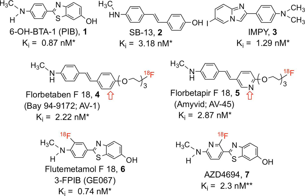
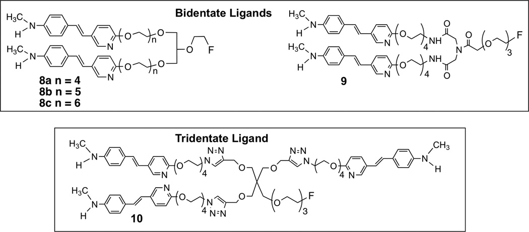
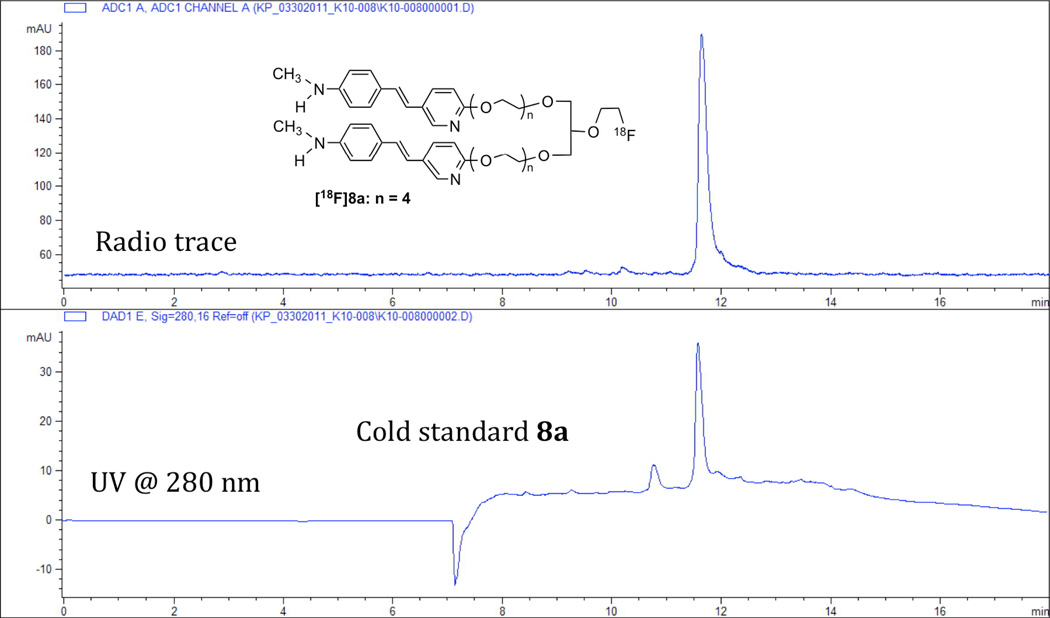
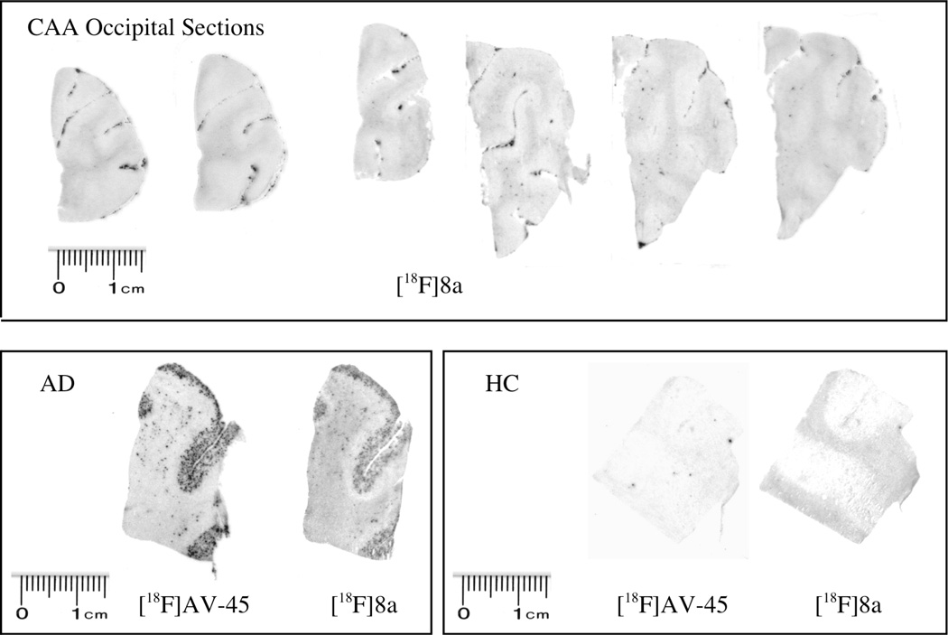
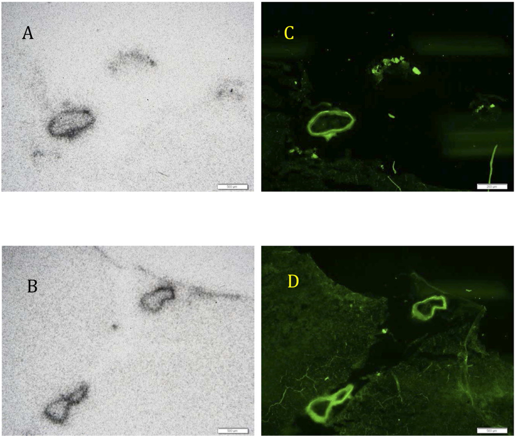
β-Amyloid plaques (Aβ plaques) in the brain are associated with cerebral amyloid angiopathy (CAA). Imaging agents that could target the Aβ plaques in the living human brain would be potentially valuable as biomarkers in patients with CAA. A new series of (18)F styrylpyridine derivatives with high molecular weights for selectively targeting Aβ plaques in the blood vessels of the brain but excluded from the brain parenchyma is reported. The styrylpyridine derivatives, 8a-c, display high binding affinities and specificity to Aβ plaques (K(i) = 2.87, 3.24, and 7.71 nM, respectively). In vitro autoradiography of [(18)F]8a shows labeling of β-amyloid plaques associated with blood vessel walls in human brain sections of subjects with CAA and also in the tissue of AD brain sections. The results suggest that [(18)F]8a may be a useful PET imaging agent for selectively detecting Aβ plaques associated with cerebral vessels in the living human brain.


Figures

Figure 1
Figure 1
The chemical structures and binding affinities of imaging agents targeting Aβ plaques in the brain are listed. All of these agents are small, neutral molecules showing high binding affinity and the ability to penetrate intact blood-brain barrier in vivo (previously published values *, **).
Figure 2
Figure 2
Multi-dentate (bidentate and tridentate) ligands, 8a, 8b, 8c, 9 and 10, based on styrylpyridine cores aiming at multiple binding sites within the repeated β-sheet structure are shown. These novel ligands are designed to bind to the Aβ aggregates via a divalent or a trivalent attachment (binding) sites.
Figure 3
Figure 3
HPLC profiles of [18F]8a and co-injected cold standard 8a on a reversed phase Gemini C18 column (250 × 4.6 mm) with the following gradient and a flow rate of 1 mL/min: 0 – 2 minutes 100% ammonium format buffer (10 mM); 2 – 5 minutes ammonium formate buffer 100% - 30%, ACN 0% – 70%; 5 –10 minutes ammonium formate buffer 30% - 0%, ACN 70% – 100%; 10 – 15 minutes 0% –100% ammonium formate buffer, 100% - 0% ACN; 15 – 18 minutes 100% ammonium formate buffer.
Figure 4
Figure 4
In the upper panel the autoradiography of postmortem human brain sections of occipital samples from cerebral amyloid angiopathy (CAA) using the new bivalent ligand, [18F]8a, showed a highly distinctive labeling at the blood vessels. Other brain parenchymal regions displayed very little labeling, which suggests that there was a selective accumulation of Aβ plaques in the vessel walls in this CAA patient. The lower panel shows brain sections of AD and healthy control (HC) subjects using either [18F]AV-45 ([18F]5) or [18F]8a. The AD brain section showed highly significant labeling of Aβ plaques, while both tracers showed little or no binding in the HC sections.
Figure 5
Figure 5
Autoradiography of postmortem human brain samples of cerebral amyloid angiopathy (CAA) using [18F]8a (A and B), which displayed excellent labeling of Aβ aggregates of the blood vessel walls. There was prominent labeling of major blood vessels only in the CAA brain sections. It is apparent that the parenchymal brain tissue had no Aβ plaques, therefore no labeling was observed. When the neighboring sections were labeled with thioflavin S (C and D), the fluorescent images displayed a matching staining of blood vessel walls comparable to the images from the autoradiography of [18F]8a. The results strongly suggest that [18F]8a labeled the Aβ aggregates of the blood vessel walls. The white bars represent a scale of 500 µm.
Scheme 1
Scheme 1
Ragent and conditions: (a) (Boc)2O, H2O, 35 °C; (b) NaH, CH3I, DMF, rt; (c) triethylene glycol, CsCO3, DMF, 150 °C; (d) K2CO3, Bu4NBr, Pd(OAc)2. DMF, 60 °C; (e) TsCl, Et3N, DMAP, DCM, rt; (f) diethylene glycol ditosylate, NaH, DMF, rt; (g) DHP, PPTS, EtOH, rt; (h)K2CO3.Bu4NI, Bu4NOAc, ACN; (i) NH3, CH3OH.
Scheme 2
Scheme 2
Ragent and conditions: (a) NaH, 2 eq. 15, DMF, rt; (b)PPTS, EtOH; (c) NaH, 2-(2-bromoethoxy)-tetrahydro-2H-pyran, DMF, rt; (d) TsCl, Et3N, DMAP, DCM, rt; (e) TBAF, THP, 70°C; (f) TFA, rt.
Scheme 3
Scheme 3
Ragent and conditions: (a)N2CHCO2C2H5, BF3 Et2O, DCM, 0 °C, (b)NBS PPh3, DCM, 0 °C, (c)NaOH, THF, rt; (d)HOBt, EDCI, DIPEA, ditert-butyl iminodiacetate, DMF rt; (e) TFA, rt; (f) NaN3, DMF, 60°C; (g)PPh3, H2O, THF, 60°C; (h) DCC, DMAP, DCM, rt; (i) TBAF, THF, 70 °C.
Scheme 4
Scheme 4
Ragent and conditions: (a)40% NaOH, DMSO, rt; (b) NaH, tri ethylene glycol di(p-toluenesulfonate), DMF, rt; (c) TBAF, THF, 70 °C; (e)sodium ascorbate, CuSO4, t-BuOH, H2O, rt; (f) TFA, rt.
Scheme 5
Scheme 5
Ragent and conditions: (a) Na[18F]floride, K[2,2,2]/K2CO3; (b) 6N HCl

Similar articles

Cited by

References

    1. Soontornniyomkij V, Choi C, Pomakian J, Vinters HV. High-definition characterization of cerebral beta-amyloid angiopathy in Alzheimer's disease. Hum. Pathol. 2010;41:1601–1608. - PMC - PubMed
    1. Fisher M, French S, Ji P, Kim RC. Cerebral microbleeds in the elderly: a pathological analysis. Stroke. 2010;41:2782–2785. - PMC - PubMed
    1. Cordonnier C. Brain microbleeds: more evidence, but still a clinical dilemma. Curr. Opin. Neurol. 2011;24:69–74. - PubMed
    1. Altmann-Schneider I, Trompet S, de Craen AJ, van Es AC, Jukema JW, Stott DJ, Sattar N, Westendorp RG, van Buchem MA, van der Grond J. Cerebral microbleeds are predictive of mortality in the elderly. Stroke. 2011;42:638–644. - PubMed
    1. Poels MM, Ikram MA, van der Lugt A, Hofman A, Krestin GP, Breteler MM, Vernooij MW. Incidence of cerebral microbleeds in the general population: the rotterdam scan study. Stroke. 2011;42:656–661. - PubMed

Publication types

MeSH terms

Substances

LinkOut - more resources