. 2012 Apr;18(7-8):816-27.
doi: 10.1089/ten.TEA.2011.0267. Epub 2011 Dec 9.

Gas-foaming calcium phosphate cement scaffold encapsulating human umbilical cord stem cells

Affiliations

Gas-foaming calcium phosphate cement scaffold encapsulating human umbilical cord stem cells

Wenchuan Chen et al. Tissue Eng Part A. 2012 Apr.

Abstract

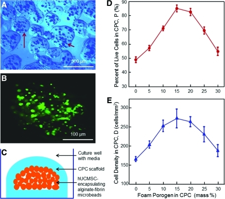
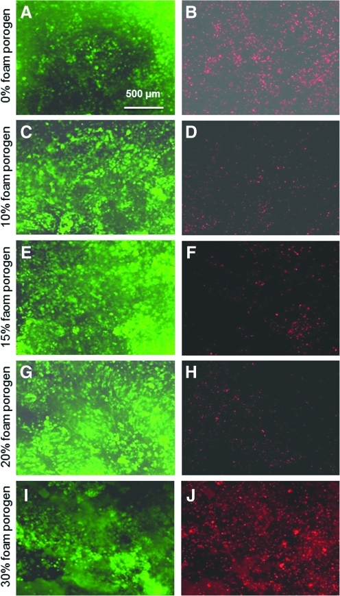
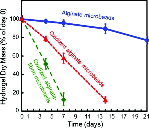
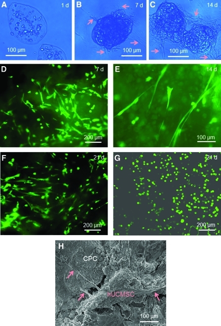
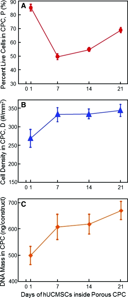
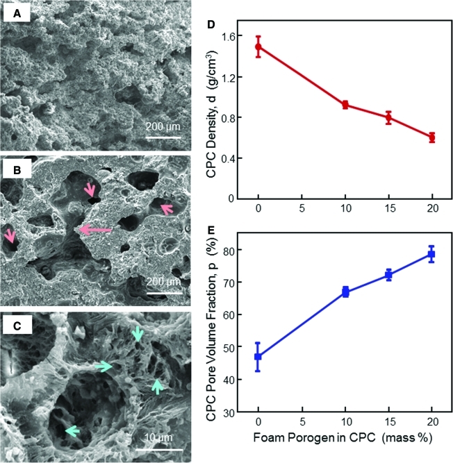
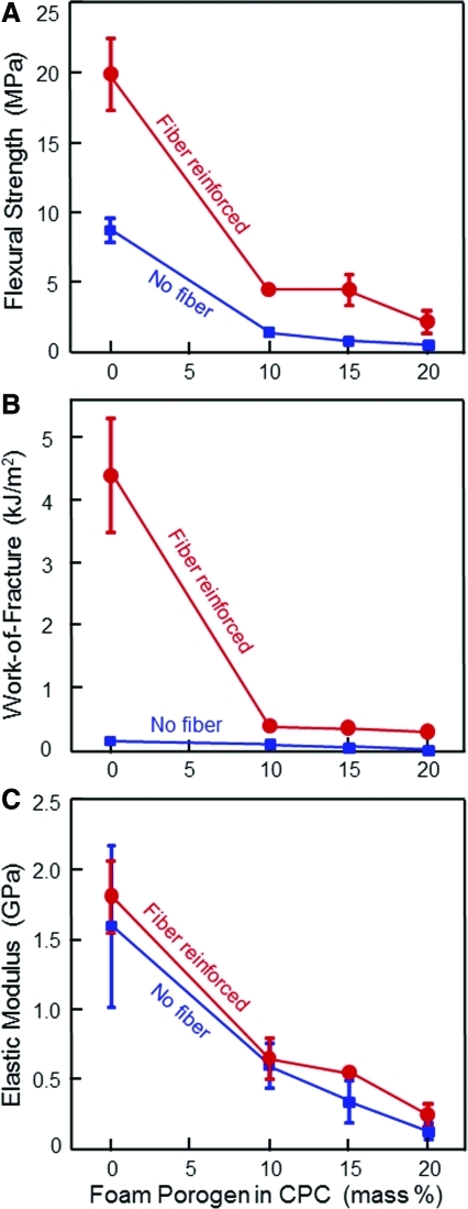
Tissue engineering approaches are promising to meet the increasing need for bone regeneration. Calcium phosphate cement (CPC) can be injected and self-set to form a scaffold with excellent osteoconductivity. The objectives of this study were to develop a macroporous CPC-chitosan-fiber construct containing alginate-fibrin microbeads encapsulating human umbilical cord mesenchymal stem cells (hUCMSCs) and to investigate hUCMSC release from the degrading microbeads and proliferation inside the porous CPC construct. The hUCMSC-encapsulated microbeads were completely wrapped inside the CPC paste, with the gas-foaming porogen creating macropores in CPC to provide for access to culture media. Increasing the porogen content in CPC significantly increased the cell viability, from 49% of live cells in CPC with 0% porogen to 86% of live cells in CPC with 15% porogen. The alginate-fibrin microbeads started to degrade and release the cells inside CPC at 7 days. The released cells started to proliferate inside the macroporous CPC construct. The live cell number inside CPC increased from 270 cells/mm(2) at 1 day to 350 cells/mm(2) at 21 days. The pore volume fraction of CPC increased from 46.8% to 78.4% using the gas-foaming method, with macropore sizes of approximately 100 to 400 μm. The strength of the CPC-chitosan-fiber scaffold at 15% porogen was 3.8 MPa, which approximated the reported 3.5 MPa for cancellous bone. In conclusion, a novel gas-foaming macroporous CPC construct containing degradable alginate-fibrin microbeads was developed that encapsulated hUCMSCs. The cells had good viability while wrapped inside the porous CPC construct. The degradable microbeads in CPC quickly released the cells, which proliferated over time inside the porous CPC. Self-setting, strong CPC with alginate-fibrin microbeads for stem cell delivery is promising for bone tissue engineering applications.


Figures

FIG. 1.
FIG. 1.
(A) Optical photo of human umbilical cord mesenchymal stem cell (hUCMSC)-encapsulating alginate–fibrin microbeads. The long arrow indicates the boundary of a microbead. The short arrow indicates the cells inside the microbeads. (B) Live/dead image of an as-fabricated microbead. Live cells were stained green, which indicates that the encapsulated cells were mostly alive. (C) Schematic of hUCMSC-encapsulating microbeads inside calcium phosphate cement (CPC) scaffold immersed in culture media in a culture well. and Effects of gas-foam porogen mass fraction in CPC on the percentage of live cells (D) and live cell density (E). Each value is the mean of five measurements, with the error showing one standard deviation (mean±standard deviation (sd); n=5). Color images available online at www.liebertonline.com/tea
FIG. 2.
FIG. 2.
Live/dead stained photos of cells inside CPC. Live cells were stained green and dead cells were stained red. Labels on the left side indicate the porogen mass fraction in CPC. The number of live cells increased, and dead cells decreased, when the porogen was increased from 0% to 10%, 15%, and 20%. Color images available online at www.liebertonline.com/tea
FIG. 3.
FIG. 3.
Microbead degradation rates. Each value is mean±sd; n=5. Three types of microbeads were measured: alginate microbeads, oxidized alginate microbeads, and oxidized alginate-fibrin microbeads. The dry mass at 1, 4, 7, 14 and 21 days was divided by the original mass at 0 days to obtain the percentage of mass remaining at each time period. Color images available online at www.liebertonline.com/tea
FIG. 4.
FIG. 4.
Alginate–fibrin microbeads degradation and cell release. (A-C) Optical photos at 1, 7, and 14 days. A blue filter was used to enhance the contrast of the microbeads. Arrows in B and C indicate cell release from the microbeads. (D-G) Live/dead staining images of the bottom CPC surface. (D) At 7 days, some cells showed as green dots, indicating that they were still encapsulated in the fragments of microbeads. Other cells had a spread, spindled morphology, indicating that they were released and attached to CPC. (E) The spread morphology at 14 days. (F) At 21 days, the number of attached cells increased. (G) For comparison, alginate microbeads without fibrin did not degrade, with the cells remaining as green dots, and did not spread at 21 days. (H) Scanning electron microscopy (SEM) micrograph of released cells attaching to the CPC bottom surface. Arrows indicate the cytoplasmic extensions of the cells. Color images available online at www.liebertonline.com/tea
FIG. 5.
FIG. 5.
Effect of time on hUCMSCs inside CPC. Each CPC contained the same 15% porogen. (A) Percentage of live cells, (B) live cell density, and (C) DNA mass of the hUCMSCs. There was a decrease in the percentage of live cells in the first week, but cell proliferation increased the percentage of live cells as well as the live cell density and the DNA mass of the hUCMSCs over time. Each value is mean±sd; n=5. Color images available online at www.liebertonline.com/tea
FIG. 6.
FIG. 6.
Gas-foaming CPC porosity. SEM micrograph at (A) 0%, (B) 10%, and (C) 20% foam porogen. An example of macropore interconnection is indicated by the long arrow in (B). The short arrows indicate openings inside macropores. Numerous micropores in CPC are shown in (C). (D, E) Effects of gas-foaming porogen on CPC density and porosity. The purpose was to measure the porosity in CPC matrix, so the specimens contained no fibers. When the foam agent was increased from 0% to 20%, the pore volume fraction in CPC increased from 46.8% (intrinsic microporosity in CPC) to 78.4% (microporosity+macroporosity). Each value is mean±sd; n=5. Color images available online at www.liebertonline.com/tea
FIG. 7.
FIG. 7.
Effect of foam porogen content on CPC mechanical properties: (A) Flexural strength, (B) work-of-fracture, and (C) elastic modulus. For the group with fibers, suture fibers at a length of 3 mm and a volume fraction of 20% were used in CPC. Each value is mean±sd; n=5. Fiber reinforcement greatly increased the CPC mechanical properties. Color images available online at www.liebertonline.com/tea

Similar articles

Cited by

References

    1. Mao J.J. Vunjak-Novakovic G. Mikos A.G. Atala A. Regenerative Medicine: Translational Approaches and Tissue Engineering. Boston, MA: Artech House; 2007.
    1. Russias J. Saiz E. Deville S. Gryn K. Liu G. Tomsia A.P. Fabrication and in vitro characterization of three-dimensional organic/inorganic scaffolds by robocasting. J Biomed Mater Res A. 2007;83:434. - PubMed
    1. Bodde E.W. Boerman O.C. Russel F.G. Mikos A.G. Spauwen P.H. Jansen J.A. The kinetic and biological activity of different loaded rhBMP-2 calcium phosphate cement implants in rats. J Biomed Mater Res A. 2008;87:780. - PubMed
    1. Atala A. Engineering organs. Curr Opin Biotechnol. 2009;20:575. - PubMed
    1. Bohner M. Design of ceramic-based cements and putties for bone graft substitution. Eur Cell Mater. 2010;20:1. - PubMed

Publication types

Substances