. 2012 Feb 29;86(2):45.
doi: 10.1095/biolreprod.111.095752. Print 2012 Feb.

Spata22, a novel vertebrate-specific gene, is required for meiotic progress in mouse germ cells

Affiliations

Spata22, a novel vertebrate-specific gene, is required for meiotic progress in mouse germ cells

Sophie La Salle et al. Biol Reprod. .

Abstract

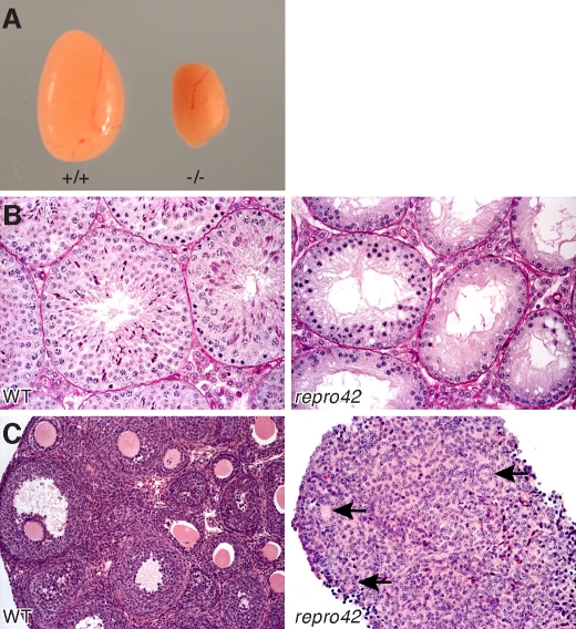
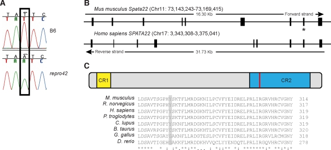
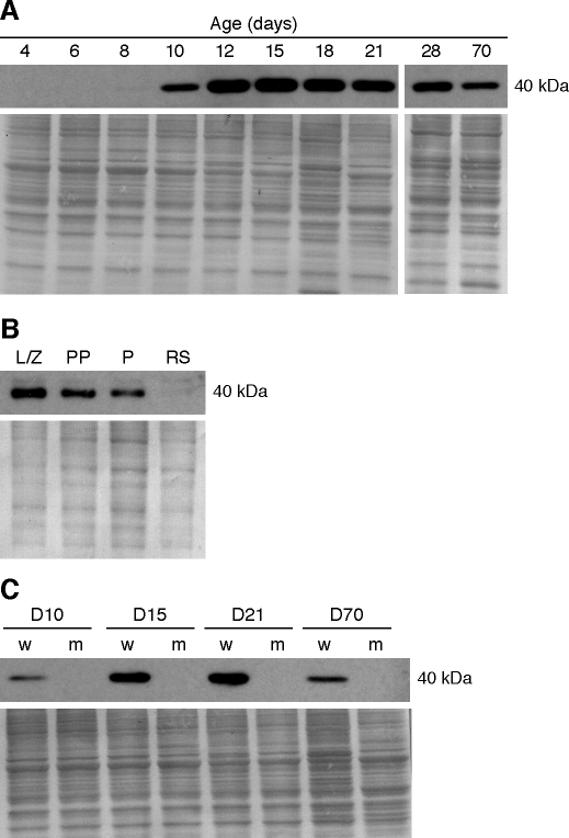
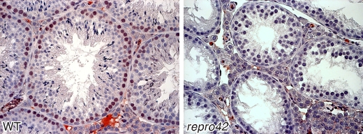
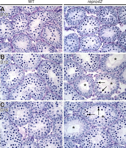
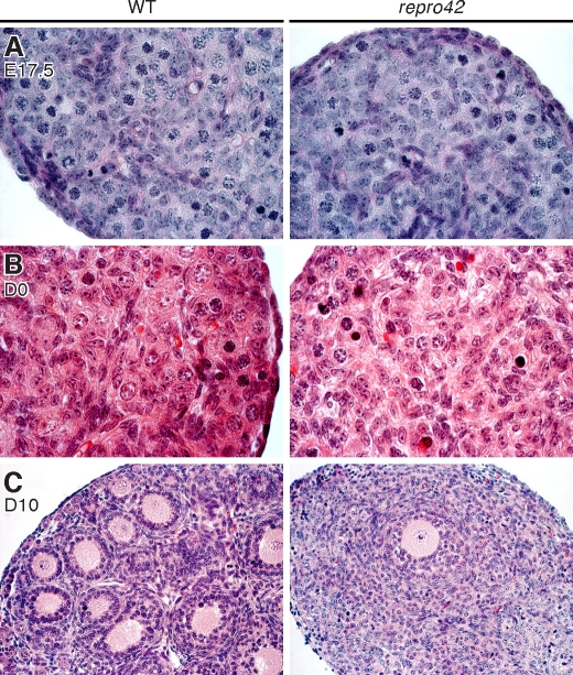
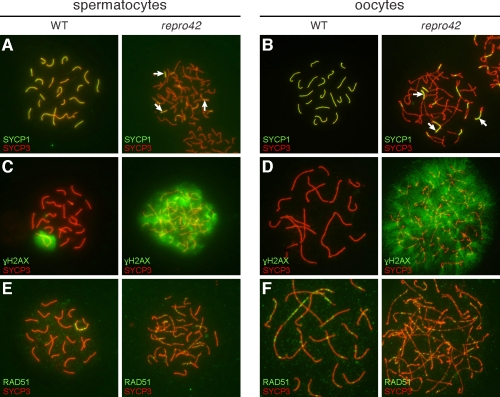
The N-ethyl-N-nitrosourea-induced repro42 mutation, identified by a forward genetics strategy, causes both male and female infertility, with no other apparent phenotypes. Positional cloning led to the discovery of a nonsense mutation in Spata22, a hitherto uncharacterized gene conserved among bony vertebrates. Expression of both transcript and protein is restricted predominantly to germ cells of both sexes. Germ cells of repro42 mutant mice express Spata22 transcript, but not SPATA22 protein. Gametogenesis is profoundly affected by the mutation, and germ cells in repro42 mutant mice do not progress beyond early meiotic prophase, with subsequent germ cell loss in both males and females. The Spata22 gene is essential for one or more key events of early meiotic prophase, as homologous chromosomes of mutant germ cells do not achieve normal synapsis or repair meiotic DNA double-strand breaks. The repro42 mutation thus identifies a novel mammalian germ cell-specific gene required for meiotic progression.


Figures

FIG. 1.
FIG. 1.
Absence of mature germ cells in adult repro42 mutant mice. A) Homozygous repro42 testes (right) are significantly smaller than those of wild-type littermate controls (left). B) Spermatogonia, spermatocytes, and spermatids are observed in testis sections from adult wild-type males (left), whereas only spermatogonia and spermatocytes up to the zygotene stage are visible in repro42 mutant testis sections (right). C) In adult wild-type females, all stages of follicular development are observed (left), whereas only degenerated follicles (black arrows) are present in repro42 mutant ovaries (right). Original magnification ×400.
FIG. 2.
FIG. 2.
A mutation in the Spata22 gene causes the repro42 phenotype. A) Sequencing of homozygous repro42 mutant DNA identified a T-to-A transversion in exon 8 of Spata22. The mutation, marked by an open box, is not present in control B6 DNA. B) Genomic structure of the mouse Spata22 and human SPATA22 genes. The exon containing the mutation, which results in a premature stop codon, is indicated by a star. C) A multiple sequence alignment performed with ClustalW2 identified two conserved regions (CR) in SPATA22. CR1 is located in the proximal end, whereas CR2 spans ∼100 amino acids and is closer to the C-termini. The identified mutation in mouse, situated at amino acid position 275, is located in CR2. A portion of the amino acid sequence of SPATA22 CR2 in eight different species is shown at the bottom. Fully conserved amino acids are marked by asterisks (*), colons (:) indicate amino acids with strongly similar physiochemical properties, and periods (.) indicate amino acids with weakly similar physiochemical properties. The position of the mutated tyrosine is marked by a red line (top) or by a pale gray box (bottom). The name of the species and the corresponding position of the last amino acid illustrated are indicated on the left and right, respectively.
FIG. 3.
FIG. 3.
Spata22 transcripts are primarily expressed in gonads in a germ cell-specific manner. A) Diagram of postulated Spata22 transcripts. The Spata22 gene is predicted to encode two transcripts: Spata22-001, which contains nine exons, and Spata22-002, which contains an additional 10th exon shared with Olfr20. The position of the primers used in the RT-PCR analysis is indicated by arrowheads for the common forward primer, an open arrow for the reverse primer for Spata22-001, and a black arrow for the reverse primer for Spata22-002. The exon containing the mutation is indicated by a star. Adapted from the Spata22 gene summary of Ensembl Genome Browser (ENSMUSG00000069825). B) Expression of Spata22 in RNA extracted from developmental series of testes collected between Postnatal Day 0 to adulthood (70 dpp). Spata22-001 was detected at all time points tested and appeared to be the sole transcript in the testis. C) Spata22 transcripts were amplified by RT-PCR in wild-type (w) and repro42 mutant (m) testes at different developmental stages. D, postnatal day. D) RT-PCR amplified Spata22 transcripts primarily in newborn (0) and 5-dpp ovaries. E) Spata22 expression was detected in meiotic and postmeiotic germ cells, but not in an extract of RNA prepared from KitW/KitW-v testes, which are devoid of differentiating germ cells. L/Z, leptotene/zygotene spermatocytes; PP, prepubertal pachytene spermatocytes; P, adult pachytene spermatocytes; RS, round spermatids; Wv, KitW/KitW-v. F) Spata22 expression in adult mice is predominantly in the testis, but also detected in liver and ES cells. Int., intestine; H20, water control.
FIG. 4.
FIG. 4.
SPATA22 protein is predominantly expressed in spermatocytes, but is absent in repro42 mutant testes. In each panel, the immunoblot results are presented at the top, and below is the corresponding stained membrane that confirms consistent protein loading and transfer. A) Immunoblotting of SPATA22 in protein extracts prepared from a developmental series of postnatal testes. SPATA22 is undetectable between 4 and 6 dpp and is faintly visible at 8 dpp, but can be readily detected from 10 dpp onwards. B) Protein extracts prepared from enriched populations of male germ cells were analyzed by Western blot. SPATA22 is present in spermatocytes but not in round spermatids. L/Z, leptotene/zygotene spermatocytes; PP, prepubertal pachytene spermatocytes; P, adult pachytene spermatocytes; RS, round spermatids. C) Analysis of protein extracts prepared from wild-type (w) and repro42 mutant (m) testes. The mutant testes lack SPATA22 detected by this antibody. D, postnatal day. The molecular weight (kDa) of SPATA22 is indicated on the right of each immunoblot.
FIG. 5.
FIG. 5.
SPATA22 protein is detected in spermatocytes and is absent in repro42 mutant testes. The brown stain in the left panel reveals expression of SPATA22 in peripheral germ cells of seminiferous tubule cross sections from wild-type (WT) adult testes, and the right panel shows absence of expression in germ cells of repro42 mutant testes. Original magnification ×400.
FIG. 6.
FIG. 6.
Meiotic arrest and loss of spermatocytes by 12 dpp in repro42 mutant testes. A) At 10 dpp, histological differences are not detectable in cross sections of repro42 mutant testes (right) when compared to cross sections of wild-type (WT) testes (left). B) At 12 dpp, spermatogonia and spermatocytes up to the pachytene stage are visible in wild-type testes (left), but differences in germ cell content become visible in mutant testes (right), where spermatocytes with condensed nuclei (arrows) and tubules with stage IV arrest (stars) are observed. C) By 15 dpp, repro42 mutant testes exhibit more spermatocytes with condensed nuclei and an increased number of tubules with stage IV arrest (right) compared to wild-type littermate controls (left). D, postnatal day. Original magnification ×400.
FIG. 7.
FIG. 7.
Meiotic arrest and loss of female germ cells in repro42 mutant ovaries after birth. A) Histology of wild-type (WT) (left) and repro42 mutant (right) ovaries at 17.5 dpc reveals the presence of numerous meiotic germ cells in mutant ovaries. Original magnification ×1000. B) Histological analysis of wild-type (left) and repro42 mutant (right) newborn (D0) ovaries reveals loss of germ cells in mutant ovaries. Original magnification ×1000. C) In 10-dpp ovaries, multiple primary and secondary follicles are visible in cross sections from wild-type mice (left), but only a few follicles remain in cross sections of repro42 mutant mice. Original magnification ×400.
FIG. 8.
FIG. 8.
Synapsis and DNA repair are impaired in repro42 mutant oocytes and spermatocytes. A, B) Colocalization of SYCP1 and SYCP3 in wild-type (WT) (left) and repro42 mutant (right) spermatocytes (A) and oocytes (B). The extent of synapsis is reflected by the pattern of yellow color representing colabeling with anti-SYCP1 (green) and anti-SYCP3 (red), detecting proteins of the CE and LEs of the SC, respectively. C, D) Labeling of γH2AX and SYCP3 in wild-type (left) and repro42 mutant (right) spermatocytes (C) and oocytes (D). The pattern of γH2AX (green) reflects DSBs and repair; the pattern of SYCP3 (red) reveals the pachytene stage of the wild-type germ cells and prepachytene of the repro42 mutant germ cells. Persistence of γH2AX suggests DSBs are induced but not repaired in mutant germ cells. E, F) Colocalization of RAD51 and SYCP3 in wild-type (left) and repro42 mutant (right) spermatocytes (E) and oocytes (F). RAD51 (green) accumulates at sites of DSBs, frequently colocalizing with SYCP3 (red), and is an early marker of meiotic recombination. In wild-type spermatocytes it is restricted to the sex chromosomes in the pachytene stage (left panel), but is found associated with autosomal chromosomes in the mutant spermatocytes. Original magnification ×630.

Comment in

Similar articles

Cited by

References

    1. Matzuk MM, Lamb DJ. The biology of infertility: research advances and clinical challenges. Nat Med 2008; 14: 1197 1213 - PMC - PubMed
    1. Costa Y, Cooke HJ. Dissecting the mammalian synaptonemal complex using targeted mutations. Chromosome Res 2007; 15: 579 589 - PubMed
    1. de Boer E, Heyting C. The diverse roles of transverse filaments of synaptonemal complexes in meiosis. Chromosoma 2006; 115: 220 234 - PubMed
    1. Page SL, Hawley RS. The genetics and molecular biology of the synaptonemal complex. Annu Rev Cell Dev Biol 2004; 20: 525 558 - PubMed
    1. Cohen PE, Pollack SE, Pollard JW. Genetic analysis of chromosome pairing, recombination, and cell cycle control during first meiotic prophase in mammals. Endocr Rev 2006; 27: 398 426 - PubMed

Publication types

MeSH terms