

Estrogen induces apoptosis in estrogen deprivation-resistant breast cancer through stress responses as identified by global gene expression across time
- PMID: 22011582
- PMCID: PMC3223472
- DOI: 10.1073/pnas.1115188108
Estrogen induces apoptosis in estrogen deprivation-resistant breast cancer through stress responses as identified by global gene expression across time
Abstract
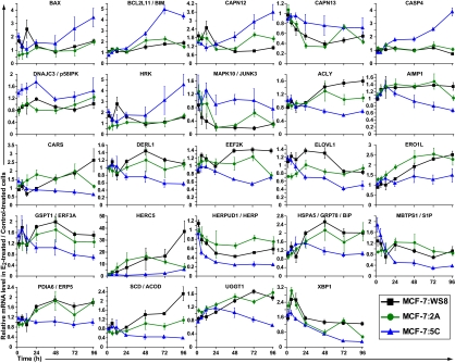
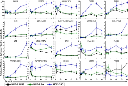
In laboratory studies, acquired resistance to long-term antihormonal therapy in breast cancer evolves through two phases over 5 y. Phase I develops within 1 y, and tumor growth occurs with either 17β-estradiol (E(2)) or tamoxifen. Phase II resistance develops after 5 y of therapy, and tamoxifen still stimulates growth; however, E(2) paradoxically induces apoptosis. This finding is the basis for the clinical use of estrogen to treat advanced antihormone-resistant breast cancer. We interrogated E(2)-induced apoptosis by analysis of gene expression across time (2-96 h) in MCF-7 cell variants that were estrogen-dependent (WS8) or resistant to estrogen deprivation and refractory (2A) or sensitive (5C) to E(2)-induced apoptosis. We developed a method termed differential area under the curve analysis that identified genes uniquely regulated by E(2) in 5C cells compared with both WS8 and 2A cells and hence, were associated with E(2)-induced apoptosis. Estrogen signaling, endoplasmic reticulum stress (ERS), and inflammatory response genes were overrepresented among the 5C-specific genes. The identified ERS genes indicated that E(2) inhibited protein folding, translation, and fatty acid synthesis. Meanwhile, the ERS-associated apoptotic genes Bcl-2 interacting mediator of cell death (BIM; BCL2L11) and caspase-4 (CASP4), among others, were induced. Evaluation of a caspase peptide inhibitor panel showed that the CASP4 inhibitor z-LEVD-fmk was the most active at blocking E(2)-induced apoptosis. Furthermore, z-LEVD-fmk completely prevented poly (ADP-ribose) polymerase (PARP) cleavage, E(2)-inhibited growth, and apoptotic morphology. The up-regulated proinflammatory genes included IL, IFN, and arachidonic acid-related genes. Functional testing showed that arachidonic acid and E(2) interacted to superadditively induce apoptosis. Therefore, these data indicate that E(2) induced apoptosis through ERS and inflammatory responses in advanced antihormone-resistant breast cancer.
Conflict of interest statement
The authors declare no conflict of interest.
Figures


References
-
- Dodds EC, Goldberg L, Lawson W, Robinson R. Estrogenic activity of certain synthetic compounds. Nature. 1938;141:247–248.
-
- Robson JM, Schönberg A. Oestrous reactions including mating produced by triphenylethylene. Nature. 1937;140:196.
-
- Haddow A. David A. Karnofsky memorial lecture. Thoughts on chemical therapy. Cancer. 1970;26:737–754. - PubMed
-
- Lerner LJ, Jordan VC. Development of antiestrogens and their use in breast cancer: Eighth Cain memorial award lecture. Cancer Res. 1990;50:4177–4189. - PubMed
Publication types
MeSH terms
Substances
Associated data
- Actions
Grants and funding
LinkOut - more resources
Full Text Sources
Medical
Molecular Biology Databases
