

Ganglioneuroma of the sphenoid wing: a case report and literature review
- PMID: 22011737
- PMCID: PMC3663467
- DOI: 10.5414/np300376
Ganglioneuroma of the sphenoid wing: a case report and literature review
Abstract
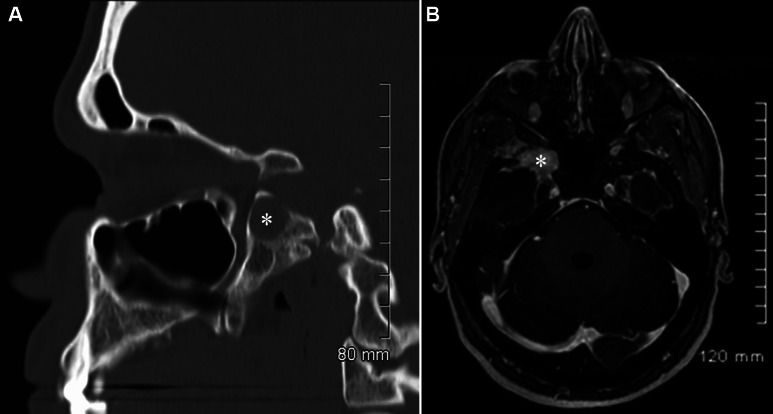
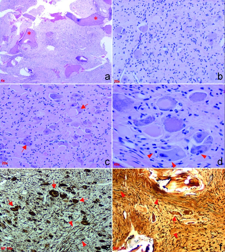
Ganglioneuromas (GNs) are well-differentiated, slow-growing, benign tumors that are quite rare and usually found in the posterior mediastinum and retroperitoneum. They are composed of ganglion and Schwann cells and their origin remains in dispute. GNs have been reported as intraosseous lesions, such as in temporal and orbital bones. There are rare reports of intracranial lesions, mostly in the pituitary fossa. Most GN patients are children and are clinically asymptomatic. Diagnosis of GN requires histopathologic evaluation since no specific clinical or radiologic diagnostic features have been identified. We report the case of a 35-year-old man with recurrent sinusitis whose radiologic workup revealed a lytic right sphenoid wing lesion with microcalcifications. He underwent gross-total resection of the lesion and the pathologic findings were diagnostic of ganglioneuroma. To the best of our knowledge, this is the first reported case of sphenoid wing GN. The nature and origin of this tumor are discussed, and the GN literature is reviewed.
Figures
References
-
- Burger PC Scheithauer BW Vogel FS Surgical pathology of the nervous system and its coverings New York: Churchill Livingstone; 2002;
-
- Ghali VS Gold JE Vincent RA Cosgrove JM Malignant peripheral nerve sheath tumor arising spontaneously from retroperitoneal ganglioneuroma: a case report, review of the literature, and immunohistochemical study. Hum Pathol. 1992; 23: 72–75 doi:10.1016/0046-8177(92)90015-U - PubMed
-
- Shin JH Lee HK Khang SK Kim DW Jeong AK Ahn KJ Choi CG Suh DC Neuronal tumors of the central nervous system: radiologic findings and pathologic correlation. Radiographics. 2002; 22: 1177–1189 - PubMed
-
- Weiss SW Goldblum JR Enzinger FM Enzinger and Weiss’s soft tissue tumors. St. Louis, Mo.: Mosby/Elsevier; 2008;
-
- Mithöfer K Grabowski EF Rosenberg AE Ryan DP Mankin HJ Symptomatic ganglioneuroma of bone. A case report. J Bone Joint Surg Am. 1999; 81: 1589–1595 - PubMed
Publication types
MeSH terms
LinkOut - more resources
Full Text Sources