

doi: 10.1109/ISBI.2009.5193165.
Mitral Annulus Segmentation from Three-Dimensional Ultrasound
Affiliations
- PMID: 22011812
- PMCID: PMC3195401
- DOI: 10.1109/ISBI.2009.5193165
Item in Clipboard
Mitral Annulus Segmentation from Three-Dimensional Ultrasound
Proc IEEE Int Symp Biomed Imaging.
2009.
Abstract
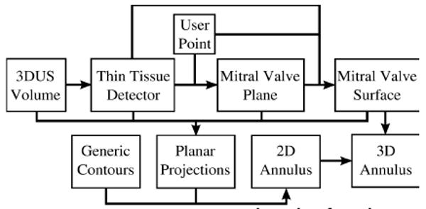
An accurate and reproducible segmentation of the mitral valve annulus from 3D ultrasound is useful to clinicians and researchers in applications such as pathology diagnosis and mitral valve modeling. Current segmentation methods, however, are based on 2D information, resulting in inaccuracies and a lack of spatial coherence. We present a segmentation algorithm which, given a single user-specified point near the center of the valve, uses max-flow and active contour methods to delineate the annulus geometry in 3D. Preliminary comparisons to manual segmentations and a sensitivity study show the algorithm is both accurate and robust.
Figures
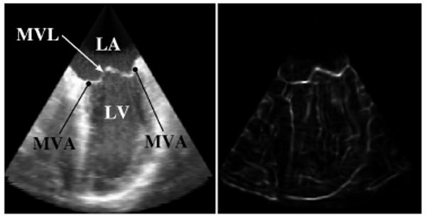
Corresponding views of a slice through a 3DUS volume showing the original intensity (left) and the TTD (right). (LA: Left Atrium; LV: Left Ventricle; MVL: Mitral Valve Leaflets; MVA: Mitral Valve Annulus)
References
-
- Salgo I, Gorman J, et al. Effect of Annular Shape on Leaflet Curvature in Reducing Mitral Leaflet Stress. Am Heart Assoc. 2002;106:711–717. - PubMed
-
- Jimenez JH, Soerensen DD, et al. Effects of a saddle shaped annulus on mitral valve function and chordal force distribution: an in vitro study. Ann Biomed Eng. 2003 Nov;31:1171–81. - PubMed
-
- Fabricius AM, Walther T, et al. Three-dimensional echocardiography for planning of mitral valve surgery: current applicability? Ann Thorac Surg. 2004 Aug;78:575–8. - PubMed
-
- Lim KH, Yeo JH, et al. Three-dimensional asymmetrical modeling of the mitral valve: a finite element study with dynamic boundaries. J Heart Valve Dis. 2005 May;14:386–92. - PubMed
-
- Lei Z, Xin Y, et al. Three Dimensional Reconstruction and Dynamic Analysis of Mitral Annular Based on Connected Equi-Length Curve Angle Chain. In: Zhang D, editor. ICMB. Vol. 4901. LNCS; 2007. pp. 298–306.
Grants and funding
LinkOut - more resources
Full Text Sources