

Fat-forming variant of solitary fibrous tumour of the pleura: CT findings
- PMID: 22011822
- PMCID: PMC3473706
- DOI: 10.1259/bjr/68692634
Fat-forming variant of solitary fibrous tumour of the pleura: CT findings
Abstract
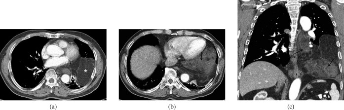
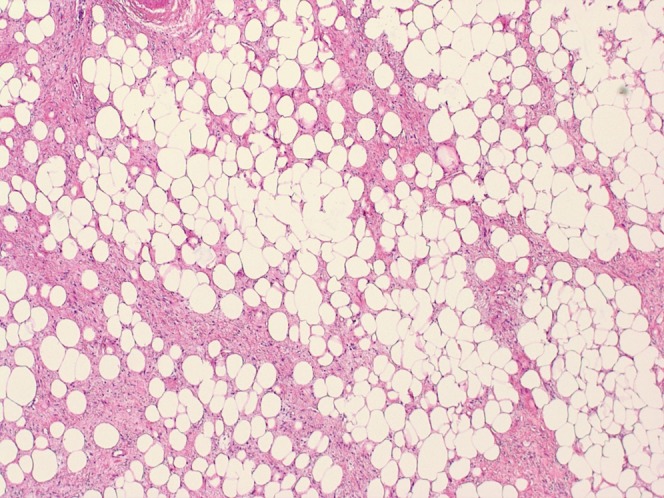
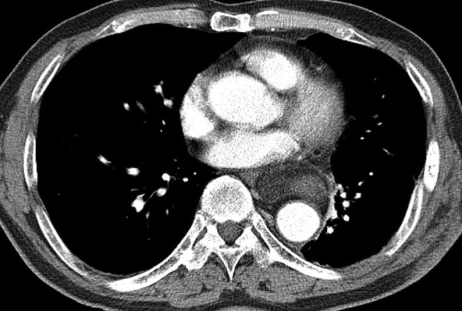
The fat-forming variant of solitary fibrous tumour (SFT) was previously called lipomatous haemangiopericytoma and is a rare variant of solitary fibrous tumour. It predominantly occurs in the deep soft tissues of the retroperitoneum and thigh. Only a handful of cases involving the perineum, spine, thoracic wall and pelvic cavity have been reported in the radiological literature and the fat-forming variant of SFT involving the pleura has not been previously reported. Herein, we report the CT findings of a case of the fat-forming variant of SFT involving the pleura that was treated by excision. Chest CT showed a large lobulated heterogeneous fatty mass with a multifocal enhancing soft-tissue component in the left lower hemithorax. Although rare, the fat-forming variant of SFT of the pleura should be added to the differential diagnosis of fat-containing pleural soft-tissue tumours.
Figures
References
-
- Gengler C, Guillou L. Solitary fibrous tumour and haemangiopericytoma: evolution of a concept. Histopathology 2006;48:63–74 - PubMed
-
- Guillou L, Gebhard S, Coindre JM. Lipomatous hemangiopericytoma: a fat-containing variant of solitary fibrous tumor? Clinicopathologic, immunohistochemical, and ultrastructural analysis of a series in favor of a unifying concept. Hum Pathol 2000;31:1108–15 - PubMed
-
- Amonkar GP, Deshpande JR, Kandalkar BM. An unusual lipomatous hemangiopericytoma. J Postgrad Med 2006;52:71–2 - PubMed
-
- Liu X, Zhang HY, Bu H, Meng GZ, Zhang Z, Ke Q. Fat-forming variant of solitary fibrous tumor of the mediastinum. Chin Med J 2007;120:1029–32 - PubMed
-
- Yamazaki K, Eyden BP. Pulmonary lipomatous hemangiopericytoma: report of a rare tumor and comparison with solitary fibrous tumour. Ultrastruct Pathol 2007;31:51–61 - PubMed
