. 2012 Mar;29(3):770-81.
doi: 10.1007/s11095-011-0601-8. Epub 2011 Oct 20.

Lapatinib distribution in HER2 overexpressing experimental brain metastases of breast cancer

Affiliations

Lapatinib distribution in HER2 overexpressing experimental brain metastases of breast cancer

Kunal S Taskar et al. Pharm Res. 2012 Mar.

Abstract

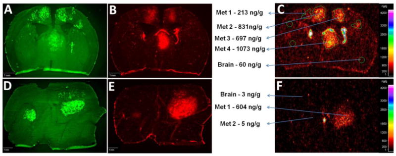
Purpose: Lapatinib, a small molecule EGFR/HER2 inhibitor, partially inhibits the outgrowth of HER2+ brain metastases in preclinical models and in a subset of CNS lesions in clinical trials of HER2+ breast cancer. We investigated the ability of lapatinib to reach therapeutic concentrations in the CNS following (14)C-lapatinib administration (100 mg/kg p.o. or 10 mg/kg, i.v.) to mice with MDA-MD-231-BR-HER2 brain metastases of breast cancer.

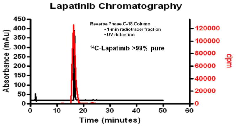
Methods: Drug concentrations were determined at differing times after administration by quantitative autoradiography and chromatography.

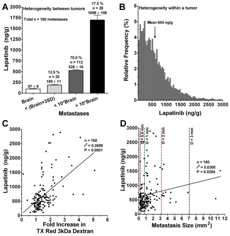
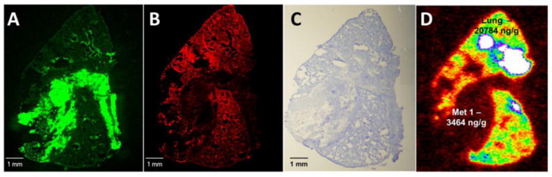
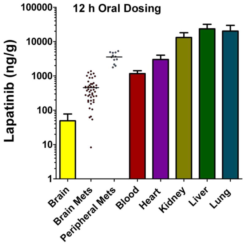
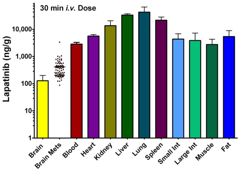
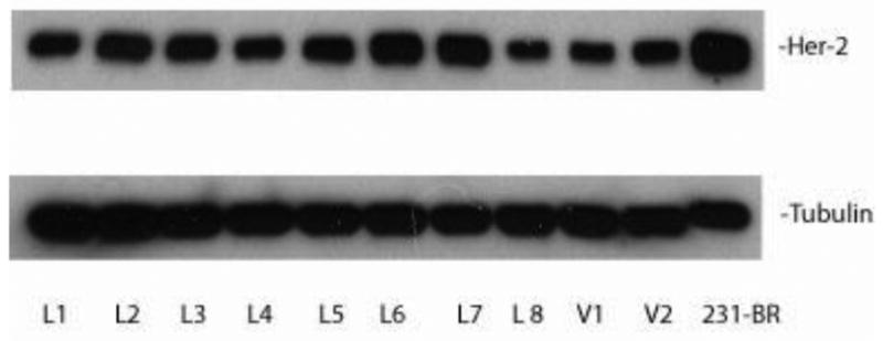
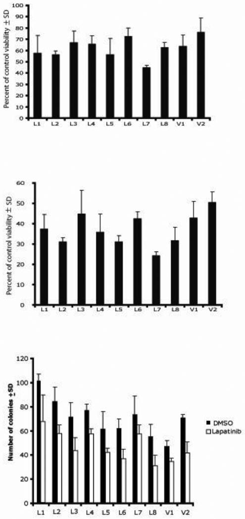
Results: (14)C-Lapatinib concentration varied among brain metastases and correlated with altered blood-tumor barrier permeability. On average, brain metastasis concentration was 7-9-fold greater than surrounding brain tissue at 2 and 12 h after oral administration. However, average lapatinib concentration in brain metastases was still only 10-20% of those in peripheral metastases. Only in a subset of brain lesions (17%) did lapatinib concentration approach that of systemic metastases. No evidence was found of lapatinib resistance in tumor cells cultured ex vivo from treated brains.

Conclusions: Results show that lapatinib distribution to brain metastases of breast cancer is partially restricted and blood-tumor barrier permeability is a key component of lapatinib therapeutic efficacy which varies between tumors.


Figures

Figure 1
Figure 1
14C-Lapatinib in brain and brain metastases at 2 (a–c) and 12 (d–f) hours following 14C-lapatinib administration (100 mg/kg, p.o.) in mice with 231-BR-HER2 tumors. Representative coronal brain sections showing green EGFP metastasis fluorescence (a,d), Texas Red 3kD dextran fluorescence (b,e), and 14C-lapatinib radioactivity (c,f) from an animal with 231-BR-HER2 brain metastases that received 14C-lapatinib for 2 (a–c) or 12 (d–f) hours. Met = metastasis.
Figure 2
Figure 2
Distribution of 14C-lapatinib between (a) and within (b) brain metastases, as well as relation of 14C-lapatinib concentration to metastasis size (c) and barrier permeability (d). Data are for animals (n=6-8) at 2 hours after oral administration of 100 mg/kg 14C-lapatinib. Similar results were found at 12 h. (a) Brain metastasis 14C-lapatinib concentrations were divided into three mutually exclusive groups: comparable to brain (<mean brain concentration + 3 × SD), <10-fold higher relative to brain, and >10-fold higher relative to brain. Bars represent group average concentration ± SEM. The percentage of metastases within each group is listed above the bar. (b) Distribution of 14C-lapatinib concentration within a single representative brain metastasis (Met-1 in Figure 1f). Each bar represents the relative frequency of concentration within small regions (pixel = 25 ×25 μm) of the tumor. Although the average concentration for the metastasis was 604 ng/g, individual regions within the metastasis varied from 30 to >4300 ng/g. (c,d) Points represent data for individual brain metastases. Color dotted lines denote average metastasis diameter (d) in mm. The Pearson correlation is shown as a solid line and was statistically significant (P<0.05).
Figure 3
Figure 3
14C-Lapatinib in lung (a–d) metastases at 12 hour after 14C-lapatinib administration (100 mg/kg, oral). Representative sections showing lung metastasis EGFP fluorescence (a), Texas Red 3kDa dextran fluorescence (b), cresyl violet staining (c) and 14C-autoradiography (d) from an animal with 231-BR-HER2 metastases that received 14C-lapatinib at 12 hours prior to euthanasia. Matching metastases were also measured in brain from the same animal.
Figure 4
Figure 4
14C-Lapatinib concentrations in brain and peripheral metastases, as well as blood and other tissues, at 12 hours after 14C-lapatinib administration (100 mg/kg, oral) in immunocompromised mice with 231-BR-HER2 metastases. Bars represent mean ± SD for n=5 animals. Lapatinib concentration (y-axis) is shown in log scale. Points represent concentrations in individual metastases; mean concentration is shown by a line.
Figure 5
Figure 5
Radiochromatography of 14C-radioactivity in plasma at 30 min after intravenous administration of 10 mg/kg 14C-lapatinib. Radiolabeled lapatinib was found to be >98% pure and co-eluted with cold lapatinib as confirmed with HPLC.
Figure 6
Figure 6
14C-Lapatinib distribution at 30 min after i.v 14C-lapatinib administration (10 mg/kg) in immunocompromised mice with 231-BR-HER2 metastases. Bars represent mean ± SD for n=3 animals. Lapatinib concentration (y-axis) is shown in log scale. Points represent concentrations in individual metastases; mean concentration is shown by a line.
Figure 7
Figure 7
HER2 expression is maintained upon ex vivo culture of lapatinib-treated brain metastatic tumor cells. 231-BR-HER2 cells were injected into immunocompromised mice, treated with either 100 mg/kg lapatinib twice daily or vehicle. At necropsy, tumor cells were cultured ex vivo from brains of lapatinib-treated (L) or vehicle-treated (V) mice. The HER2 expression of lysates of each culture was determined by western blot, using α-tubulin as a loading control.
Figure 8
Figure 8
Ex vivo cultures of lapatinib-treated 231-BR-HER2 cells remain sensitive to lapatinib in vitro. (a-b) Cultures described in Fig. 7 were assayed for viability after 72 hours of lapatinib treatment (8 μM lapatinib, a; 10 μM lapatinib, b) by MTT assay. Data represent the mean ± SD of triplicate cultures. (c) Clonogenic assays of the ex vivo cultures in 4 μM lapatinib.

References

    1. Hynesand NE, Lane HA. ERBB receptors and cancer: the complexity of targeted inhibitors. Nat Rev Cancer. 2005;5:341–354. - PubMed
    1. Lin N, Bellon J, Winer E. CNS metastases in breast cancer. J Clin Oncol. 2004;22:3608–3617. - PubMed
    1. Leyland-Jones B. Human epidermal growth factor receptor 2-positive breast cancer and central nervous system metastases. J Clin Oncol. 2009;27:5278–5286. - PubMed
    1. Clayton AJ, Danson S, Jolly S, Ryder WD, Burt PA, Stewart AL, Wilkinson PM, Welch RS, Magee B, Wilson G, Howell A, Wardley AM. Incidence of cerebral metastases in patients treated with trastuzumab for metastatic breast cancer. Br J Cancer. 2004;91:639–643. - PMC - PubMed
    1. Bendell JC, Domchek SM, Burstein HJ, Harris L, Younger J, Kuter I, Bunnell C, Rue M, Gelman R, Winer E. Central nervous system metastases in women who receive trastuzumab-based therapy for metastatic breast carcinoma. Cancer. 2003;97:2972–2977. - PubMed

Publication types

MeSH terms