. 2011 Dec 15;118(25):6544-52.
doi: 10.1182/blood-2010-11-317909. Epub 2011 Oct 19.

The ability of MLL to bind RUNX1 and methylate H3K4 at PU.1 regulatory regions is impaired by MDS/AML-associated RUNX1/AML1 mutations

Affiliations

The ability of MLL to bind RUNX1 and methylate H3K4 at PU.1 regulatory regions is impaired by MDS/AML-associated RUNX1/AML1 mutations

Gang Huang et al. Blood. .

Abstract

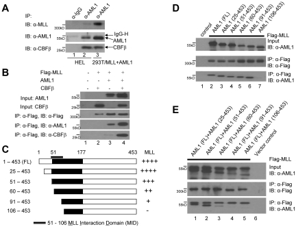
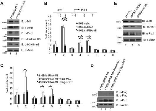
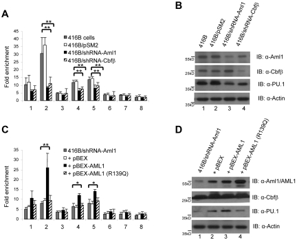
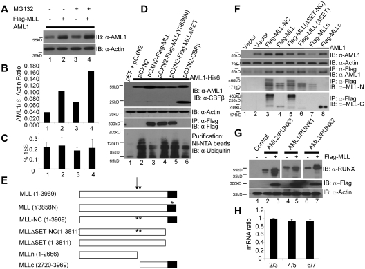
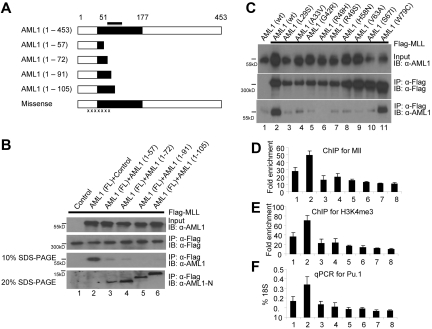
The mixed-lineage leukemia (MLL) H3K4 methyltransferase protein, and the heterodimeric RUNX1/CBFβ transcription factor complex, are critical for definitive and adult hematopoiesis, and both are frequently targeted in human acute leukemia. We identified a physical and functional interaction between RUNX1 (AML1) and MLL and show that both are required to maintain the histone lysine 4 trimethyl mark (H3K4me3) at 2 critical regulatory regions of the AML1 target gene PU.1. Similar to CBFβ, we show that MLL binds to AML1 abrogating its proteasome-dependent degradation. Furthermore, a subset of previously uncharacterized frame-shift and missense mutations at the N terminus of AML1, found in MDS and AML patients, impairs its interaction with MLL, resulting in loss of the H3K4me3 mark within PU.1 regulatory regions, and decreased PU.1 expression. The interaction between MLL and AML1 provides a mechanism for the sequence-specific binding of MLL to DNA, and identifies RUNX1 target genes as potential effectors of MLL function.


Figures

Figure 1
Figure 1
Physical interaction between MLL and AML1. (A) Interaction between the endogenous MLL and AML1 proteins in HEL cells. HEL cell nuclear lysates were used for immunoprecipitation (lane 1-2) with IgG and anti-AML1 polyclonal antibodies cross-linked with protein A sepharose beads followed by Western blot with the indicated antibodies. Nuclear lysates prepared from 293T cells that coexpress Flag tagged MLL and AML1 were used as an IP control (lane 3) with the anti-AML1 polyclonal antibodies cross-linked with protein A sepharose beads. (B) Interaction of MLL and AML1 when both are expressed in 293T cells in the absence or presence of CBFβ, followed by immunoprecipitation (IP) and Western blot. The transfections were indicated as in lane 1, vector alone; lane 2, Flag tagged MLL and CBFβ; lane 3, Flag tagged MLL and AML1; and lane 4, Flag tagged MLL, AML1, and CBFβ. Row 1 indicates AML1 input, row 2 indicates CBFβ input, row 3 indicates Flag tagged MLL IP and Western blot, row 4 indicates Western blot of AML1 with IP samples in row 3, and row 5 indicates Western blot of CBFβ with IP samples in row 3. (C) Diagram of a series of AML1 N-terminal deletion constructs: AML1 (1-453, 25-453, 51-453, 60-453, 90-453, and 106-453), and the strength of their interaction with MLL as indicated (either – or + derived from experiment in panel E). (D) Determination of the MLL interacting domain in AML1 by IP and Western blot using the N-terminal deletion constructs of AML1 shown in panel C. (E) Competitive interaction between MLL and full-length AML1 and coexpressed N-terminal deletion constructs of AML1 in 293T cells. The upper band in lanes 1-5, top panel indicate AML1 (1-453), while the lower band in lanes 1-5 indicate AML1 (25-453, 51-453, 60-453, 90-453, and 106-453). The bands (both upper and lower) in lanes 1-3, bottom panel indicate the same bands as the top panel, whereas lanes 4-5 indicate only the AML1 (1-453) band, but not the AML1 (90-453 or 106-453) bands.
Figure 2
Figure 2
Mll dictates the level of H3K4 tri-methylation at PU.1 regulatory regions. (A) Western blot of nuclear extracts from control cells (lane1) and Mll knockdown cells (lane 2) with indicated antibodies, anti-MLL, anti-AML1, anti-PU.1, anti-H3, anti-H3K4me3, and anti-actin. (B) Chromatin immunoprecipitation (ChIP) assays were performed using H3K4 tri-methylation specific antibodies and multiple primer sets to assay H3K4me3 levels at the PU.1 locus to detect the effects of shRNA-mediated knockdown of Mll in the early myeloid progenitor 416B cells. The ChIP primers localized at 1, 5′ URE; 2, 3′URE; 3, −5kb; 4, promoter; 5, +0.4 kb; 6, +6 kb; 7, +17 kb at PU.1 locus; 8, control primers at Gapdh gene locus. (C) Rescue of the H3K4me3 marks in the PU.1 URE and promoter regions after reintroduction of full-length human MLL, but not a deletion mutant form (MLL-ΔSET) that lacks a SET domain; in 416B cells that express shRNA knock down of the Mll. (D) IP or direct Western blot with indicated antibodies in established MLL and MLL-ΔSET stable expression 416B cell lines: lane 1, 416B cells that express shRNA knock down of the Mll; lane 2, 416B cells that express shRNA knock down of the Mll and overexpress Flag tagged MLL; lane 3, 416B cells that express shRNA knock down of the Mll and overexpress Flag tagged MLL-ΔSET. Row 1 indicates IP with anti-Flag beads and Western blot with Flag antibodies; row 2 indicated direct Western blot with anti-PU.1 antibodies; and row 3 indicated direct Western blot with anti-actin antibodies. (E) Western blots of nuclear extracts isolated from pLKO.1 infected control bone marrow Lin c-Kit+ cells (lane 1) and Mll knockdown cells (lane 2-3) using anti-MLL, anti-AML1, anti-PU.1, and anti-actin antibodies.
Figure 3
Figure 3
Both Aml1 and Cbfβ are required for maintaining H3K4 tri-methylation at PU.1 regulatory regions. (A) ChIP assays were performed to assess the level of H3K4 trimethylation at the PU.1 locus in 416B cells in which knockdown of either Aml1 or Cbfβ was accomplished using specific shRNAs. (B) Reduction in PU.1 levels following knockdown of either Aml1 or Cbfβ. Western blots were done using indicated antibodies (row 1, anti-AML1; row 2, anti-CBFβ; row 3, anti-PU.1) with controls (lane 1-2) and Aml1 and Cbfβ knockdown cells (lane 3-4). (C) Rescue of the H3K4me3 mark within the PU.1 URE and promoter regions following reintroduction of wild-type human AML1, but not a DNA binding mutant form of AML1 (R139Q). (D) Loss of PU.1 expression following knockdown of AML1, which could be restored following AML1 overexpression, but not AML1 (R139Q) overexpression. Western blot using indicated antibodies (row 1, anti-AML1; row 2, anti-CBFβ; row 3, anti-PU.1) with controls (lane 1-2) and AML1 (lane 3) and AML1 (R139Q; lane 4) overexpression in Aml1 knockdown cells.
Figure 4
Figure 4
MLL stabilizes AML1. (A) Stabilization of AML1 in 293T cells by coexpression of MLL or presence of MG132, a proteasome inhibitor. (B) Ratio of AML1/β-Actin is determined by densitometry based on the Western blot in Figure 4A. (C) Real-time PCR measurement of AML1 mRNA levels in experiment shown in Figure 4A. (D) Inhibition of AML1 poly-ubiquitination by coexpression of CBFβ and wild-type MLL but not MLL mutants. Lane 1, transfection control; lane 2, AML1-His6 with pCXN2 vector; lane 3, AML1-His6 with Flag-MLL; lane 4, AML1-His6 with Flag-MLL (Y3858N); lane 5, AML1-His6 with Flag-MLLΔSET; lane 6, AML1-His6 with CBFβ. Western blots were performed using the indicated antibodies (row 1, anti-AML1 and anti-CBFβ; row 2, anti-actin; row 3, IP with anti-Flag and IB with anti-Flag; row 4, His-tag AML1 were affinity purified with Ni-NTA magnetic Agarose beads and IB with anti-ubiquitin). (E) Diagram of a series of MLL deletion mutant constructs: MLL (1-3969), MLL (Y3858N), MLL-NC (1-3969), MLLΔSET-NC (1-3811), MLL ΔSET (1-3811), MLLn (1-2666), and MLLc (2720-3969). The arrows indicate 2 taspase I processing sites and the stars indicate the sites of the taspase I processing site mutations (cleavage site 1 mutation, D2666A/G2667A; cleavage site 2 mutation, D2718A/G2719A). (F) Interaction between AML1 and MLL deletion proteins. Lane 1, transfection control; lane 2, AML1 with pCXN2 vector; lane 3, AML1 with Flag-MLL-NC (noncleavable); lane 4, AML1 with Flag-MLL; lane 5, AML1 with Flag-MLLΔSET-NC; lane 6, AML1 with Flag-MLL-ΔSET; lane 7, AML1 with Flag-MLLn; and lane 8, AML1 with Flag-MLLc. Western blots were done using the indicated antibodies (row 1, anti-AML1; row 2, anti-actin; row 3, IP with anti-Flag and IB with anti-AML1; row 4, IP with anti-Flag and IB with anti-MLLn; row 5, IP with anti-Flag and IB with anti-MLLc). (G) Stabilization of AML1 family proteins (AML1, AML2, and AML3) by MLL. AML2 was expressed in lane 2 and 3. AML1 was expressed in lane 4 and 5. AML3 was expressed in lane 6 and 7. Flag-MLL was expressed in lane 3, 5, and 7. Western blots were done using indicated antibodies (row 1, anti-RUNX; row 2, anti-Flag; row 3, anti-actin). Spaces have been inserted in the top panel to indicate a repositioned gel lane. (H) AML2, AML1, and AML3 mRNA expression level ratios measured by real-time PCR assay in experiment of 4G. Lane 1, AML2 mRNA levels ratio between AML2 without MLL/AML2 with MLL; lane 2, AML1 mRNA levels ratio between AML1 without MLL/AML1 with MLL; lane 3, AML2 mRNA levels ratio between AML3 without MLL/AML3 with MLL.
Figure 5
Figure 5
The interaction between MLL and AML1 is impaired by some mutant AML1 proteins found in leukemia patients. (A) Diagram of a series of the N-terminal frame shift and misssense mutations. The black bar indicates the MID. The black area indicates the Runt-domain. The black x's indicate missense mutation of AML1 found in MID (details in Figure 5C). (B) The AML1 frame shift mutants interact with MLL and block the interaction between wild-type AML1 and MLL. Lane 1, transfection control; lane 2, AML1 with vector control; lane3, AML1 with AML1 (1-57); lane 4, AML1 with AML1 (1-72); lane 5, AML1 with AML 1(1-91); and lane 6, AML1 with AML1 (1-105). Western blots were done using indicated antibodies (row 1, anti-AML1; row 2, IP with anti-Flag and IB with anti-Flag; row 3, IP with anti-Flag, running with 10% SDS-PAGE gel and IB with anti-AML1; and row 4, IP with anti-Flag, running with 20% SDS-PAGE gel and IB with anti–AML1-N). (C) The N-terminal missense mutations of AML1 impair MLL interaction. Lane 1, transfection with pCS2 vector control and AML1; lane 2, MLL with AML1; lane 3, MLL with AML1 (L29S); lane 4, MLL with AML1 (A33V); lane 5, MLL with AML1 (G42R); lane 6, MLL with AML1 (R49H); lane 7, MLL with AML1 (R49S); lane 8, MLL with AML1 (H58N); lane 9, MLL with AML1 (V63A); lane 10, MLL with AML1 (S67I); and lane 11, MLL with AML1 (W79C). Western blots were done using indicated antibodies (row 1, anti-AML1; row 2, IP with anti-Flag and IB with anti-Flag; and row 3, IP with anti-Flag, IB with anti-AML1). (D) ChIP assay with anti-MLL antibodies and with primer set at 3′URE region of PU.1 gene (Figure 2A primer set 2) with 416B stable cell lines: 1, pBEX; 2, pBEX-AML1; 3, pBEX-AML1 (1-91); 4, pBEX-AML1 (1-105); 5, pBEX-AML1 (L29S); 6, pBEX-AML1 (H58N); 7, pBEX-AML1 (R139Q); and 8, pBEX-AML1 (R177Q). (E) ChIP assay with anti-H3K4me3 antibodies and with primer set at 3′URE region of PU.1 gene (Figure 2A primer set 2) in 416B stable cell lines in experiments 5D. (F) Real-time PCR assay for PU.1 expression in 416B stable cell lines in experiments in panels D and E.

Similar articles

Cited by

References

    1. Zhang Y, Rowley JD. Chromatin structural elements and chromosomal translocations in leukemia. DNA Repair (Amst) 2006;5(9-10):1282–1297. - PubMed
    1. Yoshida H, Kitabayashi I. Chromatin regulation by AML1 complex. Int J Hematol. 2008;87(1):19–24. - PubMed
    1. Look AT. Oncogenic transcription factors in the human acute leukemias. Science. 1997;278(5340):1059–1064. - PubMed
    1. Tkachuk DC, Kohler S, Cleary ML. Involvement of a homolog of Drosophila trithorax by 11q23 chromosomal translocations in acute leukemias. Cell. 1992;71(4):691–700. - PubMed
    1. Gu Y, Nakamura T, Alder H, et al. The t(4;11) chromosome translocation of human acute leukemias fuses the ALL-1 gene, related to Drosophila trithorax, to the AF-4 gene. Cell. 1992;71(4):701–708. - PubMed

Publication types

MeSH terms