. 2011 Nov;106(6):1023-39.
doi: 10.1007/s00395-011-0228-2. Epub 2011 Oct 20.

Cyclic nucleotide phosphodiesterase 1A: a key regulator of cardiac fibroblast activation and extracellular matrix remodeling in the heart

Affiliations

Cyclic nucleotide phosphodiesterase 1A: a key regulator of cardiac fibroblast activation and extracellular matrix remodeling in the heart

Clint L Miller et al. Basic Res Cardiol. 2011 Nov.

Abstract

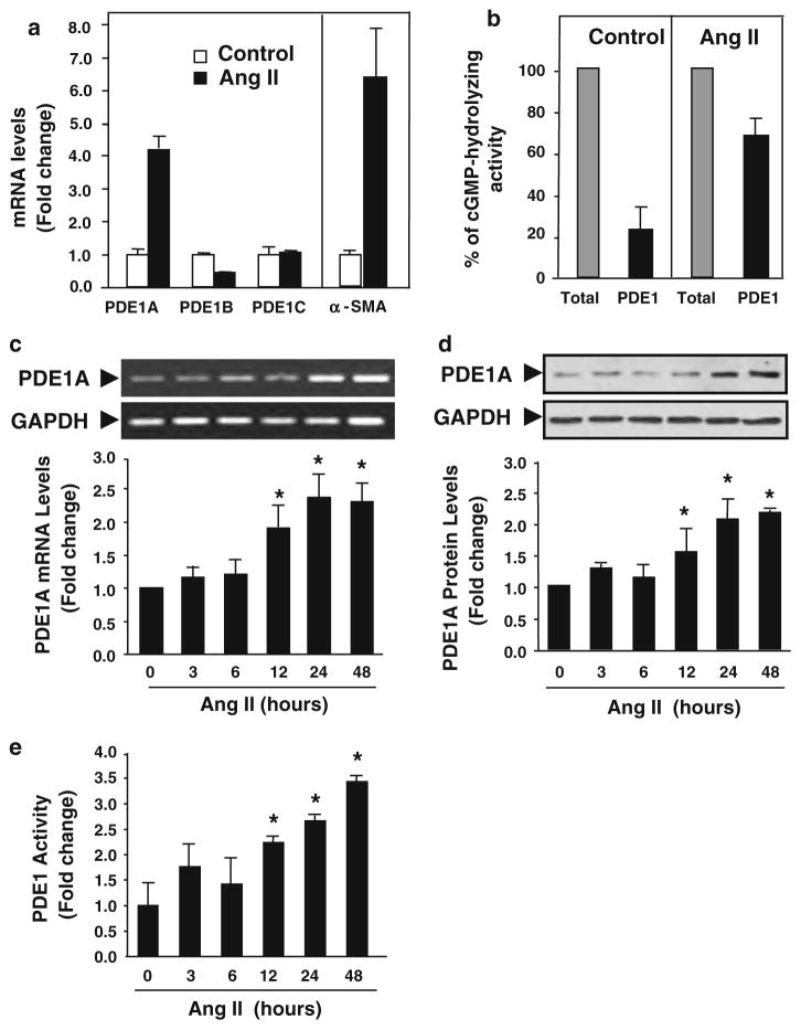
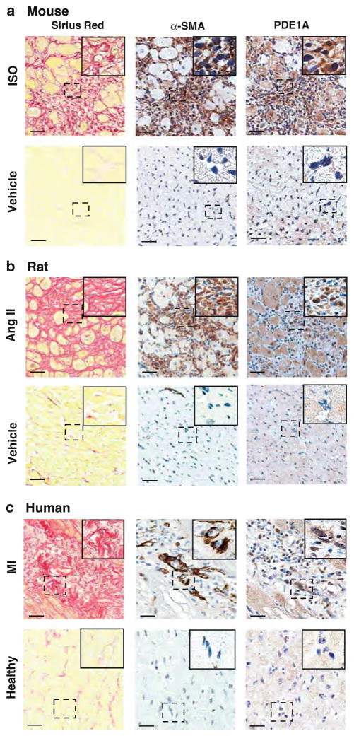
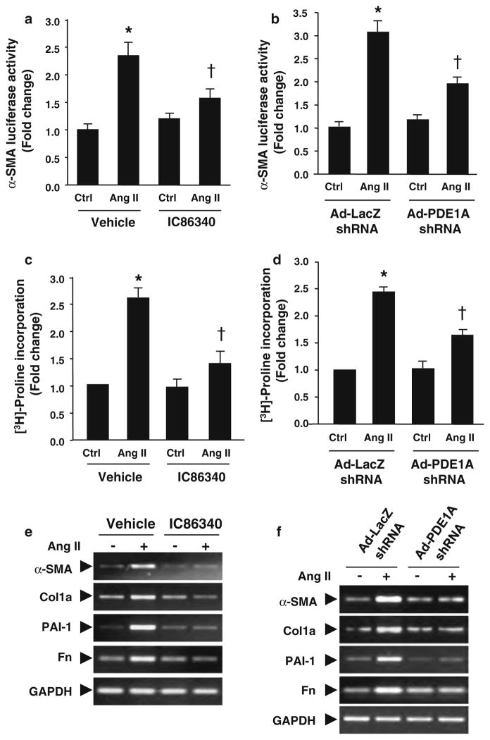
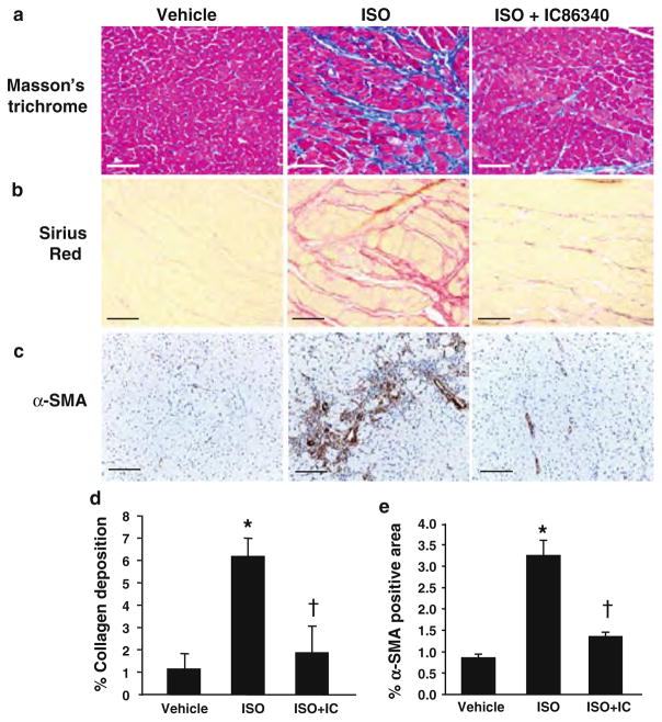
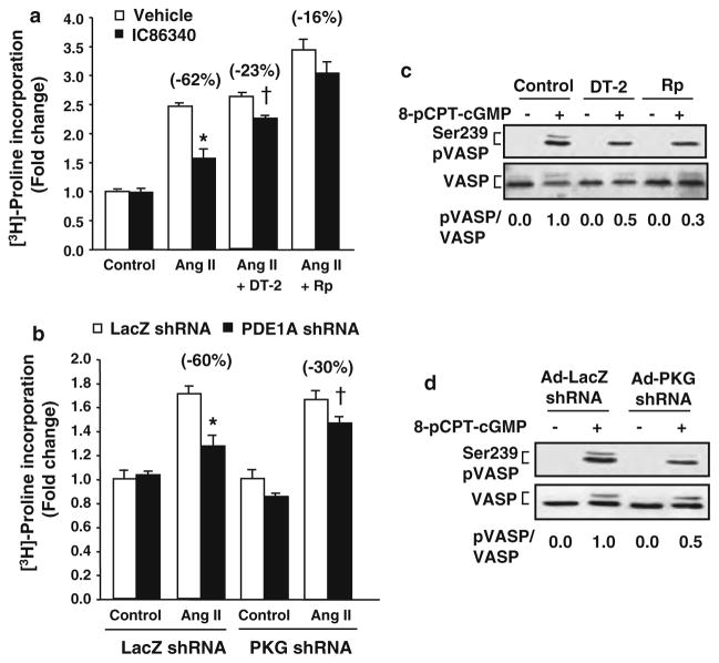
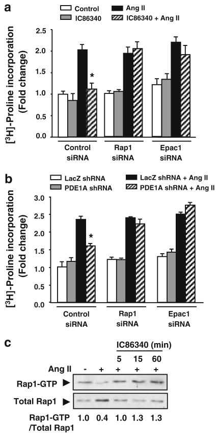
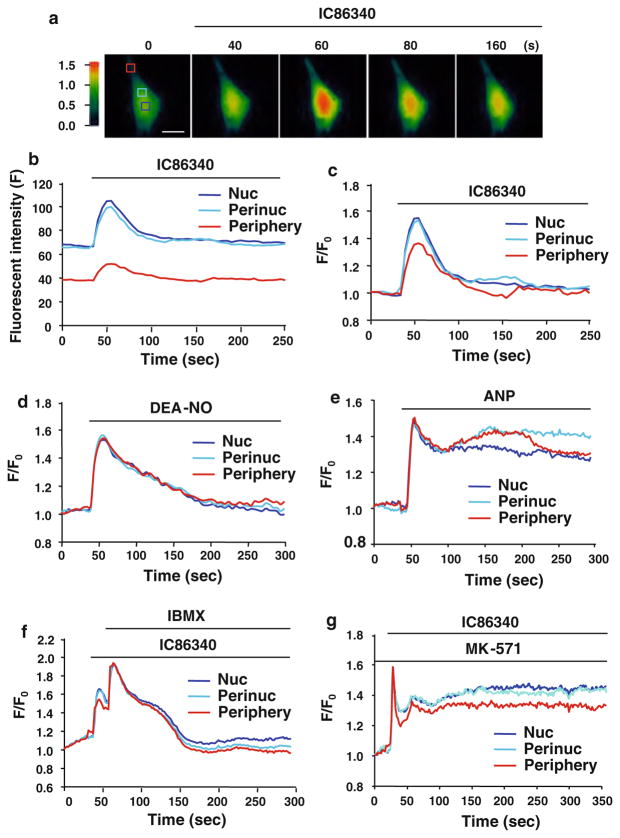
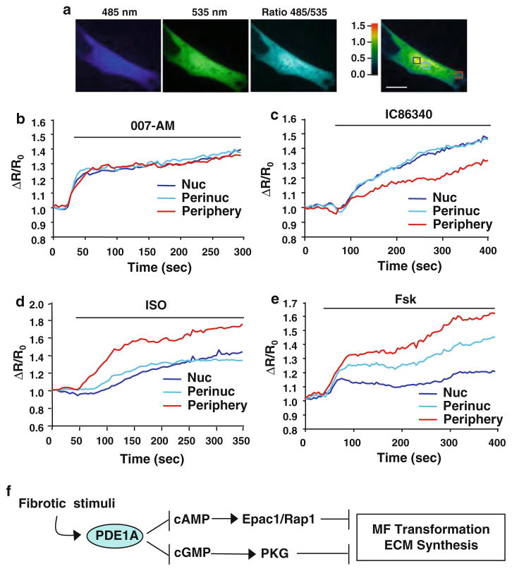
Cardiac fibroblasts become activated and differentiate to smooth muscle-like myofibroblasts in response to hypertension and myocardial infarction (MI), resulting in extracellular matrix (ECM) remodeling, scar formation and impaired cardiac function. cAMP and cGMP-dependent signaling have been implicated in cardiac fibroblast activation and ECM synthesis. Dysregulation of cyclic nucleotide phosphodiesterase (PDE) activity/expression is also associated with various diseases and several PDE inhibitors are currently available or in development for treating these pathological conditions. The objective of this study is to define and characterize the specific PDE isoform that is altered during cardiac fibroblast activation and functionally important for regulating myofibroblast activation and ECM synthesis. We have found that Ca(2+)/calmodulin-stimulated PDE1A isoform is specifically induced in activated cardiac myofibroblasts stimulated by Ang II and TGF-β in vitro as well as in vivo within fibrotic regions of mouse, rat, and human diseased hearts. Inhibition of PDE1A function via PDE1-selective inhibitor or PDE1A shRNA significantly reduced Ang II or TGF-β-induced myofibroblast activation, ECM synthesis, and pro-fibrotic gene expression in rat cardiac fibroblasts. Moreover, the PDE1 inhibitor attenuated isoproterenol-induced interstitial fibrosis in mice. Mechanistic studies revealed that PDE1A modulates unique pools of cAMP and cGMP, predominantly in perinuclear and nuclear regions of cardiac fibroblasts. Further, both cAMP-Epac-Rap1 and cGMP-PKG signaling was involved in PDE1A-mediated regulation of collagen synthesis. These results suggest that induction of PDE1A plays a critical role in cardiac fibroblast activation and cardiac fibrosis, and targeting PDE1A may lead to regression of the adverse cardiac remodeling associated with various cardiac diseases.


Figures

Fig. 1
Fig. 1
PDE1A expression and activity are increased by Ang II stimulation in rat cardiac fibroblasts. a Real-time quantitative RT-PCR results comparing the mRNA expression levels of PDE1 isoforms in control (serum-free) and Ang II stimulation conditions for 24 h in rat cardiac fibroblasts, normalized to GAPDH mRNA levels. α-SMA levels represent the conversion to cardiac myofibroblasts. Values represent mean ± SD. Results are typical from two independent experiments performed in triplicate. b cGMP PDE1 assay performed in the presence of 1 μM cGMP substrate demonstrating the effects of Ang II (100 nmol/L) stimulation (24 h) on PDE1 inhibitable (via 10 μmol/L IC86340) cGMP hydrolyzing activity in rat cardiac fibroblasts. Results are presented as the percentage of total cGMP hydrolyzing activity. Values represent mean ± SD from at least three independent experiments. c Semi-quantitative RT-PCR results showing the effects of Ang II stimulation for the indicated times on PDE1A mRNA expression relative to GAPDH mRNA levels. Values represent mean ± SD from four experiments. d Western blot showing the effects of Ang II stimulation for the indicated times on PDE1A protein levels in cardiac fibroblasts. Values represent mean ± SD from three experiments. e Ca2+-dependent PDE assay showing the effects of Ang II stimulation for various times on PDE1 cGMP hydrolyzing activity in rat cardiac fibroblasts. Values are mean ± SD of triplicates from a typical experiment and are representative of five independent experiments. *P < 0.05 versus 0 h
Fig. 2
Fig. 2
PDE1A protein levels are increased in activated cardiac myofibroblasts in fibrotic regions of mouse, rat, and human diseased hearts. a Mouse hearts subjected to 1-week ISO infusion (30 mg/kg/day) or vehicle control and stained for collagen deposition and immunostained for α-SMA and PDE1A (n = 6–8 per group). b Rat hearts subjected to 2 weeks Ang II infusion (0.7 mg/kg/day) or vehicle control and stained for collagen deposition and immunostained for α-SMA and PDE1A (n = 3 per group). c Post-MI remodeled human hearts or normal control hearts stained for collagen deposition, and immunostained for α-SMA and PDE1A. (n = 4 per group). Left panels Representative high magnification images of picro-sirius red staining depicting increased interstitial collagen deposition in mouse, rat, and human hearts. Middle panels Adjacent sections immunostained for α-SMA confirm the induction of cardiac myofibroblasts. Right panels PDE1A immunostaining depicts increased PDE1A expression in interstitial myofibroblasts in remodeled hearts. Insets, higher magnification of boxed areas. PDE1A expression is differentially localized in nuclear and cytosolic compartments in cardiac fibroblasts within the left ventricle. Scale bar = 20 μm
Fig. 3
Fig. 3
PDE1 inhibitors and PDE1A knockdown attenuates myofibroblast activation, ECM synthesis and pro-fibrotic gene expression. a Cardiac fibroblast activation assessed by α-SMA promoter luciferase activity in rat cardiac fibroblasts pre-treated with vehicle or IC86340 (30 μmol/L) prior to Ang II (100 nmol/L) stimulation for 24 h, and normalized to β-galactosidase activity. b Cardiac fibroblasts transduced with 100 MOI Ad-LacZ shRNA or Ad-PDE1A shRNA and stimulated as described above. c Collagen synthesis measured via of [3H]-proline incorporation in cardiac fibroblasts pre-treated with vehicle or IC86340 prior to Ang II stimulation for 48 h, and normalized to total protein levels. d Cardiac fibroblasts transduced with Ad-LacZ shRNA or Ad-PDE1A shRNA and stimulated as described above. Values represent mean ± SD from three independent experiments performed in triplicates. *P < 0.05 versus Ctrl (Vehicle or LacZ shRNA), †P < 0.05 versus Ang II alone. e Representative RT-PCR results showing effects of the PDE1 inhibitor IC86340 on Ang II stimulated pro-fibrotic marker gene expression, including type I collagen (Col1a), fibronectin (Fn), plasminogen activator inhibitor 1 (PAI-1), and α-SMA. Similar results were observed from at least three independent experiments. f RT-PCR results showing effects of PDE1A shRNA versus LacZ shRNA on Ang II stimulated pro-fibrotic gene expression in cardiac fibroblasts. Similar results were observed from at least two independent experiments
Fig. 4
Fig. 4
PDE1 inhibition attenuates collagen deposition and myofibroblast formation in response to ISO induced cardiac remodeling in vivo. Mice at age of 10–12 weeks were infused with 30 mg/kg/day for 1 week and treated with vehicle or IC86340 (3 mg/kg/day, i.p.). a, b Representative images of collagen deposition in left ventricle of mouse hearts depicted by Masson’s trichrome staining (a) or picro-sirius red staining (b) of vehicle control (left panel), ISO (middle panel) and ISO + IC86340 (right panel) treatment groups. Scale bar = 30 μm (Masson’s trichrome staining) or = 20 μm (picro-sirius red). c Effects of IC86340 treatment on myofibroblast formation assessed by α-SMA immunostaining. Scale bar = 20 μm. d, e Quantitated results of percentage area of picro-sirius staining in B, of background subtracted interstitial and perivascular collagen (including left and right ventricle and intraventricular septum) and percentage area of α-SMA staining in whole myocardium for each treatment group (n = 9–12). Values represent mean ± SD. *P < 0.05 versus Vehicle, †P < 0.05 versus ISO
Fig. 5
Fig. 5
PKG-I dependent effects of PDE1 inhibition on collagen synthesis. a Collagen synthesis measured via [3H]-proline incorporation in cardiac fibroblasts treated with vehicle or the PDE1 inhibitor IC86340 (30 μmol/L) in the presence of vehicle or PKG-I inhibitors, DT-2 (250 nmol/L) or Rp-8-Br-PET-cGMPS (20 μmol/L) and stimulated with Ang II (100 nmol/L) for 48 h, and normalized to total protein levels. Values represent mean ± SD from a typical experiment performed in triplicate. Similar results were observed from n = 3 independent experiments. *P < 0.05 versus Ang II + Vehicle, †P < 0.05 versus Ang II + DT-2 + Vehicle. b Collagen synthesis in cardiac fibroblasts transduced with adenovirus expressing LacZ shRNA (Ad-LacZ shRNA) or PKG I (Ad-PKG I shRNA) prior to IC86340 treatment and Ang II stimulation. Values represent mean ± SD from a typical experiment performed in triplicate. Similar results were observed from n = 3 independent experiments. *P < 0.05 versus Ang II + LacZ shRNA, †P < 0.05 versus Ang II + PKG shRNA + LacZ shRNA. c, d Effects of PKG I inhibitors and PKG I shRNA on PKG-dependent Ser239 VASP phosphorylation. Cardiac fibroblasts were pre-treated with DT-2 or Rp-8-Br-PET-cGMPS for 30 min or transduced with Ad-LacZ shRNA or Ad-PKG shRNA for 48 h prior to stimulation with 8-pCPT-cGMP (100 μmol/L) for 30 min. Western blots were performed using a monoclonal antibody recognizing phosphorylated Ser239 VASP or polyclonal antibody against total VASP. The ratio of phosphorylated VASP to total VASP shown below each blot was determined via densitometry in a linear range
Fig. 6
Fig. 6
Role of Epac1-Rap1 signaling in mediating effects of PDE1 inhibition. a Collagen synthesis measured via [3H]-proline incorporation in cardiac fibroblasts transfected with siRNA against non-targeting control, Rap1, or Epac1 followed by treatment with vehicle or the PDE1 inhibitor IC86340 (30 μmol/L) and Ang II (100 nmol/L) stimulation for 48 h, and normalized to total protein levels. b Collagen synthesis in cardiac fibroblasts transduced with Ad-Lacz shRNA or PDE1A shRNA for 48 h followed by Ang II stimulation, and normalized to total protein levels. Values represent mean ± SD of triplicates from n = 3 independent experiments. *P < 0.05 versus Ang II or LacZ shRNA + Ang II. c Rap1 activity determined using GST-RalGDS-RBD to pulldown Rap1-GTP in cardiac fibroblasts stimulated with Ang II (100 nmol/L) for 24 h prior to treatment with PDE1 inhibitor IC86340 (30 μmol/L) for the indicated times. Rap1-GTP levels shown below each blot were quantitated by densitometry in a linear range and normalized to total Rap1 levels. Values (normalized to serum-free control) are averaged from two independent experiments
Fig. 7
Fig. 7
Effects of PDE1 inhibition on spatial and temporal cGMP changes in activated cardiac fibroblasts. a Pseudo-colored images depict the relative PDE1 inhibition-induced cGMP changes measured by GFP-based cGMP sensor δ-FlincG in primary cardiac fibroblasts activated in the presence of 5% serum. Activated cardiac fibroblasts transduced with adenovirus δ-FlincG were treated with the PDE1 inhibitor IC86340 (15 μmol/L) and the fluorescence intensities (excitation at 485 nm and emission at 510 nm) were captured. Boxes denote regions of interests in the nucleus (Nuc), perinucleus (Perinuc), and peripheral compartments. Scale bar = 25 μm. b Fluorescence intensity traces from the above boxed regions after IC86340 treatment with background fluorescence subtracted. c Background subtracted intensity traces normalized to the fluorescence intensity at 0 s (ΔF/F0) for each compartment. Results are typical from n = 10. d Activated cardiac fibroblasts expressing Ad-δ-FlincG and treated with the NO donor DEA-NONOate (2.5 nmol/L) elicits a transient cGMP response in each region of interests. Results are typical from n = 12. e ANP (30 nmol/L) treatment results in sustained cGMP response in each region of interests. Results are typical from n = 8. f Effects of global PDE inhibition via IBMX (100 μmol/L) on PDE1 inhibitor induced transient cGMP response. Results are typical from n = 8. g Effects of cGMP efflux transporter MRP4/5 inhibitor MK-571 (20 μmol/L) on PDE1 inhibitor induced transient cGMP response. Results are typical from n = 6
Fig. 8
Fig. 8
Effects of PDE1 inhibition on spatial and temporal cAMP changes in activated cardiac fibroblasts. a Primary rat cardiac fibroblasts (activated in the presence of 5% serum) were co-transfected with Epac1-camps and the nuclear targeted Epac1-H30-NLS which resulted in a relatively uniform distribution of the cAMP sensor throughout the cytosol, nucleus, perinucleus, and cell periphery regions. Pseudo-colored fluorescence intensity is shown for the FRET donor, CFP (485 nm), the acceptor YFP (535 nm), corrected FRET ratio 485/535 nm. Boxes denote regions of interests in the nucleus (Nuc), perinucleus (Perinuc), and peripheral compartments. Scale bar = 10 μm. b Stimulation of activated cardiac fibroblasts with the membrane permeable Epac-selective agonist, 007-AM (1 μmol/L) generates a rapid and uniform increase in 485/535 confirming the functionality of the sensor. Data are represented as the fluorescence intensity changes in the FRET donor/acceptor ratio 485/535 normalized to the baseline ratio at 0 s (ΔR/R0), for each regions of interest. c Effects of the PDE1 inhibitor IC86340 (15 μmol/L) on 485/535 which elicits changes in [cAMP]i preferentially in the nucleus (Nuc) and perinucleus (Perinuc) compared to the cell periphery (Periphery). d Stimulation of activated cardiac fibroblasts with ISO (0.1 μmol/L) generates an increase in cAMP predominately in the cell periphery. e Stimulation with the AC activator Forskolin (Fsk; 10 μmol/L) elicits an increase in intracellular cAMP, differentially in the regions of interests, preferentially at the cell periphery. Results are typical from n = 6–10 per treatment. f Proposed model of PDE1A-dependent dual regulation of cAMP-Epac1-Rap1 and cGMP-PKG signaling to suppress myofibroblast (MF) transformation and ECM synthesis during pathological cardiac remodeling

Similar articles

Cited by

References

    1. Aizawa T, Wei H, Miano JM, Abe J, Berk BC, Yan C. Role of phosphodiesterase 3 in NO/cGMP-mediated antiinflammatory effects in vascular smooth muscle cells. Circ Res. 2003;93:406–413. doi: 10.1161/01.RES.0000091074.33584.F0. - DOI - PubMed
    1. Andric SA, Kostic TS, Stojilkovic SS. Contribution of multidrug resistance protein MRP5 in control of cyclic guanosine 5′-monophosphate intracellular signaling in anterior pituitary cells. Endocrinology. 2006;147:3435–3445. doi: 10.1210/en.2006-0091. - DOI - PubMed
    1. Batchelor AM, Bartus K, Reynell C, Constantinou S, Halvey EJ, Held KF, Dostmann WR, Vernon J, Garthwaite J. Exquisite sensitivity to subsecond, picomolar nitric oxide transients conferred on cells by guanylyl cyclase-coupled receptors. Proc Natl Acad Sci USA. 2010;107:22060–22065. doi: 10.1073/pnas.1013147107. - DOI - PMC - PubMed
    1. Bax NA, van Oorschot AA, Maas S, Braun J, van Tuyn J, de Vries AA, Groot AC, Goumans MJ. In vitro epithelial-to-mesenchymal transformation in human adult epicardial cells is regulated by TGFbeta-signaling and WT1. Basic Res Cardiol. 2011;106:829–847. doi: 10.1007/s00395-011-0181-0. - DOI - PMC - PubMed
    1. Beavo JA, Brunton LL. Cyclic nucleotide research—still expanding after half a century. Nat Rev Mol Cell Biol. 2002;3:710–718. doi: 10.1038/nrm911. - DOI - PubMed

Publication types

MeSH terms

LinkOut - more resources