. 2011 Oct 20;2(10):e221.
doi: 10.1038/cddis.2011.104.

OGX-427 inhibits tumor progression and enhances gemcitabine chemotherapy in pancreatic cancer

Affiliations

OGX-427 inhibits tumor progression and enhances gemcitabine chemotherapy in pancreatic cancer

V Baylot et al. Cell Death Dis. .

Abstract

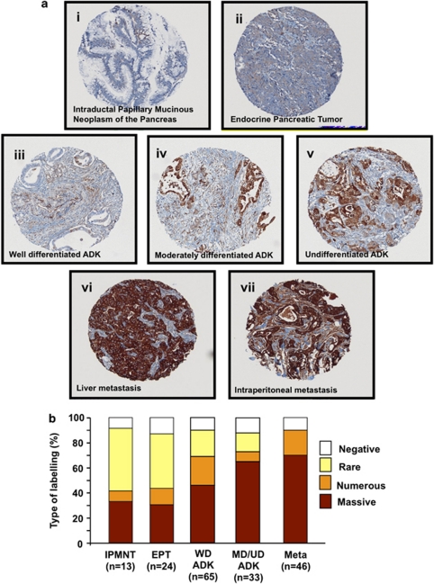
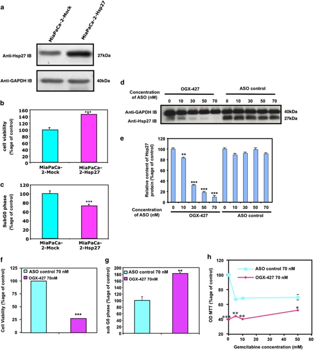
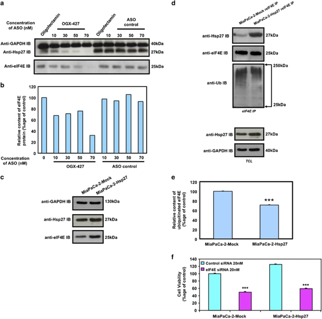
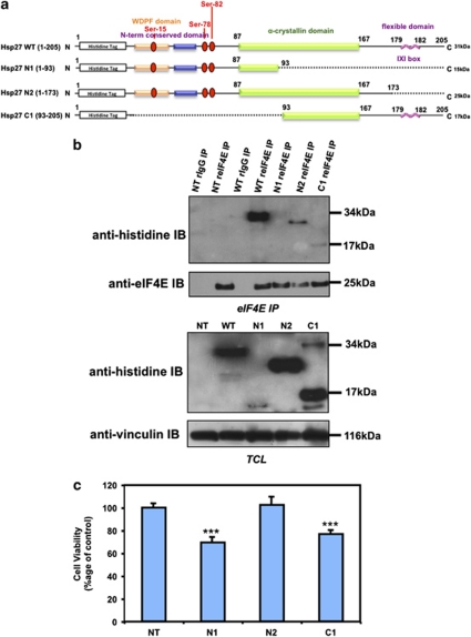
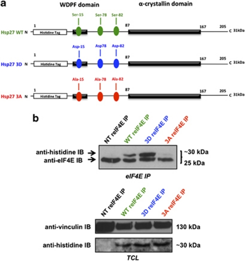
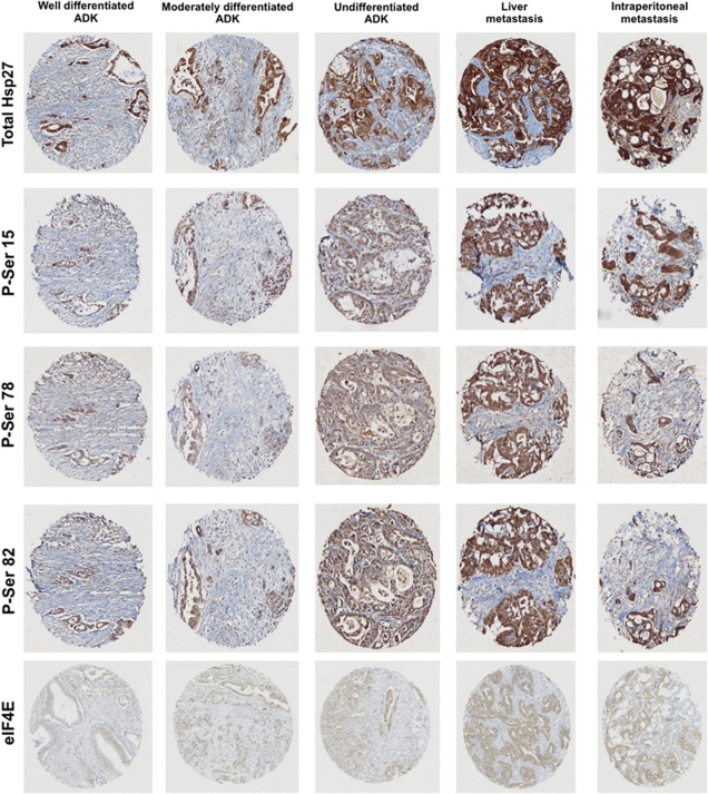
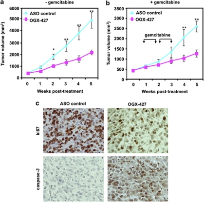
Despite many advances in oncology, almost all patients with pancreatic cancer (PC) die of the disease. Molecularly targeted agents are offering hope for their potential role in helping translate the improved activity of combination chemotherapy into improved survival. Heat shock protein 27 (Hsp27) is a chaperone implicated in several pathological processes such as cancer. Further, Hsp27 expression becomes highly upregulated in cancer cells after chemotherapy. Recently, a modified antisense oligonucleotide that is complementary to Hsp27 (OGX-427) has been developed, which inhibits Hsp27 expression and enhances drug efficacy in cancer xenograft models. Phase II clinical trials using OGX-427 in different cancers like breast, ovarian, bladder, prostate and lung are in progress in the United States and Canada. In this study, we demonstrate using TMA of 181 patients that Hsp27 expression and phosphorylation levels increase in moderately differentiated tumors to become uniformly highly expressed in metastatic samples. Using MiaPaCa-2 cells grown both in vitro and xenografted in mice, we demonstrate that OGX-427 inhibits proliferation, induces apoptosis and also enhances gemcitabine chemosensitivity via a mechanism involving the eukaryotic translation initiation factor 4E. Collectively, these findings suggest that the combination of Hsp27 knockdown with OGX-427 and chemotherapeutic agents such as gemcitabine can be a novel strategy to inhibit the progression of pancreas cancer.


Figures

Figure 1
Figure 1
Changes in Hsp27 immunostaining in human pancreas cancer TMA. (ai and aii) IPMNP and EPT: a few foci of weakly positive cancer cells are visible, but most tumor is not immunoreactive. (aiii, aiv and av) Strong immunoreactivity in WD, MD differentiated and UD ductal adenocarcinoma. (avi and avii) Sheets of uniformly and intensively reactive tumor cells characteristic of liver and intraperitoneal Meta, respectively. (b) Mean Hsp27 staining in human PC TMA. Specimens spotted on the TMA were graded from negative, rare, numerous and massive representing the range of no staining to heavy staining by visual scoring and automated quantitative image analysis by SAMBA ‘immune' software. Data from 181 samples were used to calculate average ±S.E. All comparison of stain intensity was made at × 200 magnification
Figure 2
Figure 2
Effect of Hsp27 overexpression and downregulation on chemoresistant MiaPaCa-2 cells survival and apoptosis in vitro. (a) Western blot analysis of Hsp27 and GAPDH protein levels in MiaPaCa-2 cells stably transfected with empty vector (MiaPaCa-2-Mock) or human Hsp27 (MiaPaCa-2-Hsp27). (b) MTT quantification of cell viability of MiaPaCa-2-Hsp27 and -Mock cells and (c) apoptosis assay by flow cytometry. The results are expressed in percentage to control (MiaPaCa-2-Mock). (d) Western blot analysis of Hsp27 and GAPDH protein levels in MiaPaCa-2 cells transfected with OGX-427 as compared with MiaPaCa-2 ASO control. (e) Histograms of average densitometries of Hsp27 protein levels after normalization to GAPDH protein levels by densitometry analysis in MiaPaCa-2 cells after treatment with OGX-427 or ASO control. (f) MTT quantification of cell viability of MiaPaCa-2 cells treated with OGX-427 or ASO control. (g) Flow cytometry quantification of cell percentage in sub G0 phase of MiaPaCa-2 cells treated with OGX-427 as compared with ASO control. (h) MTT quantification of cell viability of MiaPaCa-2 treated with OGX-427 or ASO control combined with different concentrations of gemcitabine. Error bars represent S.E. from three independent experiments. Statistical analysis used t test; *P≤0.05, **P≤0.01, ***P≤0.001
Figure 3
Figure 3
Hsp27 regulates eIF4E and mediates cytoprotection. (a) Western blot analysis of Hsp27, eIF4E and GAPDH protein levels in MiaPaCa-2 cells treated with OGX-427 or ASO control. (b) Histograms of average densitometries of eIF4E protein level after normalization to GAPDH protein level by densitometry analysis in MiaPaCa-2 cells after treatment with OGX-427 or ASO control. (c) Western blot analysis of Hsp27, eIF4E and GAPDH protein levels in MiaPaCa-2 cells stably transfected with empty vector (MiaPaCa-2-Mock) or human Hsp27 (MiaPaCa-2-Hsp27). (d) Western blot analysis of Hsp27, ubiquitin and eIF4E protein levels after eIF4E immunoprecipitation (IP) using rabbit anti-eIF4E antibody (reIF4E) in MiaPaCa-2-Mock and MiaPaCa-2-Hsp27 cells. Total cell lysate (TCL) represents proteins from MiaPaCa-2-Mock vs MiaPaCa-2-Hsp27 cells, extracted from cultured cells and blotted as control with Hsp27 and GAPDH antibody. (e) Histograms of average densitometries of ubiquitine protein levels after normalization to eIF4E protein levels by densitometry analysis in MiaPaCa-2 stably transfected with Hsp27 or Mock control. (f) MTT quantification of cell viability of MiaPaCa-2-Mock and MiaPaCa-2-Hsp27 cells treated with 20 nM control- or eIF4E-siRNA and gemcitabine (150 mM). Error bars represent S.E. from three independent experiments. Statistical analysis used t test; ***P≤0.001
Figure 4
Figure 4
Hsp27 association with eIF4E involves its C-terminal region. (a) Schematic representation of Hsp27 structure and deletion mutants used in this study. (b) Western blot analysis of histidine and eIF4E protein levels after eIF4E immunoprecipitation (IP) using rabbit anti-eIF4E antibody (reIF4E) or rabbit IgG as control in MiaPaCa-2 cells transiently transfected with WT Hsp27 or truncated mutant forms (N1, N2 and C1) of Hsp27. In the lower panel, western blot analysis of histidine and vinculin levels from total protein extracts (TCL). (c) MTT quantification of cell viability of MiaPaCa-2 cells transiently transfected or not with Hsp27 deletion mutants and treated with gemcitabine (150 mM). Error bars, S.E. from three independent experiments. Statistical analysis used t test; ***P≤0.001
Figure 5
Figure 5
Hsp27-eIF4E interaction depends on Hsp27 phosphorylation. (a) Schematic representation of WT and phosphorylation mutants (3D and 3A) of Hsp27 used in this study. (b) Western blot analysis of histidine and eIF4E protein levels after eIF4E immunoprecipitation (IP) using rabbit anti-eIF4E antibody (reIF4E) in MiaPaCa-2 cells transiently transfected with Hsp27 WT or 3D (constitutively phosphorylated) and 3A (constitutively dephosphorylated) vectors. In the lower panel, western blot analysis of histidine and vinculin levels from total protein extracts (TCL)
Figure 6
Figure 6
Total Hsp27, phospho-Hsp27 and eIF4E levels in PC tumors. Immunohistochemistry analysis for Hsp27, P-Hsp27 and eIF4E in human pancreas cancer tumors. Specimens from well-differentiated, moderately-differentiated, undifferentiated ADK and metastasis (liver, intraperitoneal) were stained with phospho-serine-15 (P-Ser 15), -78 (P-Ser 78) and -82 (P-Ser 82) Hsp27, total Hsp27 or eIF4E in back to back sections
Figure 7
Figure 7
Anti-cancer effect of OGX-427 on MiaPaCa-2 tumor growth and gemcitabine sensitivity in vivo. (a) and (b) MiaPaCa-2 tumor volume measurement (length × width × depth × 0.5236) in 40 nude randomly selected for treatment with OGX-427 or ASO control. When MiaPaCa-2 tumors reached 300–500 mm3, OGX-427 or ASO control were injected i.p. for 5 weeks for OGX-427 (a and b) (see Materials and Methods section). Points represent mean tumor volume in each experimental group containing 10 mice; Error bars represent S.E. Statistical analysis used t test; *P≤0.05, **P≤0.01. (c) IHC staining of MiaPaCa-2 tumor xenografts. Tumors were resected and processed for histological analysis as previously described in Materials and Methods sections. Tissue sections were stained with antibodies to ki67 and cleaved caspase-3

Similar articles

Cited by

References

    1. Van Laethem JL, Verslype C, Iovanna JL, Michl P, Conroy T, Louvet C, et al. New strategies and designs in pancreatic cancer research: consensus guidelines report from a European expert panel Ann Oncol 2011. e-pub ahead of print 1 August 2011. - PubMed
    1. Conroy T, Desseigne F, Ychou M, Bouche O, Guimbaud R, Becouarn Y, et al. FOLFIRINOX versus gemcitabine for metastatic pancreatic cancer. N Engl J Med. 2011;364:1817–1825. - PubMed
    1. Cunningham D, Chau I, Stocken DD, Valle JW, Smith D, Steward W, et al. Phase III randomized comparison of gemcitabine versus gemcitabine plus capecitabine in patients with advanced pancreatic cancer. J Clin Oncol. 2009;27:5513–5518. - PubMed
    1. Egloff AM, Grandis JR. Response to combined molecular targeting: defining the role of P-STAT3. Clin Cancer Res. 2011;17:393–395. - PMC - PubMed
    1. Wang SJ, Gao Y, Chen H, Kong R, Jiang HC, Pan SH, et al. Dihydroartemisinin inactivates NF-kappaB and potentiates the anti-tumor effect of gemcitabine on pancreatic cancer both in vitro and in vivo. Cancer Lett. 2010;293:99–108. - PubMed

Publication types

MeSH terms