Review
. 2011 Dec;92(5):921-8.
doi: 10.1007/s00253-011-3637-0. Epub 2011 Oct 20.

Murein and pseudomurein cell wall binding domains of bacteria and archaea--a comparative view

Affiliations
Review

Murein and pseudomurein cell wall binding domains of bacteria and archaea--a comparative view

Ganesh Ram R Visweswaran et al. Appl Microbiol Biotechnol. 2011 Dec.

Abstract

The cell wall, a major barrier protecting cells from their environment, is an essential compartment of both bacteria and archaea. It protects the organism from internal turgor pressure and gives a defined shape to the cell. The cell wall serves also as an anchoring surface for various proteins and acts as an adhesion platform for bacteriophages. The walls of bacteria and archaea are mostly composed of murein and pseudomurein, respectively. Cell wall binding domains play a crucial role in the non-covalent attachment of proteins to cell walls. Here, we give an overview of the similarities and differences in the biochemical and functional properties of the two major murein and pseudomurein cell wall binding domains, i.e., the Lysin Motif (LysM) domain (Pfam PF01476) and the pseudomurein binding (PMB) domain (Pfam PF09373) of bacteria and archaea, respectively.


Figures

Fig. 1
Fig. 1
Hidden Markov model (HMM) showing the consensus sequence of the LysM (Pfam database entry PF01476) and the PMB motifs (Pfam database entry PF09373). The HMM logo pictures were generated separately from the Pfam database for a the LysM motifs and b the PMB motifs, using their respective Pfam accession numbers. The X-axis indicates the relative entropy, and the contribution of each amino acid residue is shown on the Y-axis. Numbers on the X-axis indicate the position of the amino acid residue in the total length of the motif. The size of the amino acid residues on Y-axis is directly proportional to their conservation in the consensus sequence
Fig. 2
Fig. 2
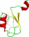
Crystal structure of the LysM motif of YkuD from B. subtilis. The figure was generated using PyMOL (The PyMOL Molecular Graphics System, Version 1.2r3pre, Schrödinger, LLC) and PDB entry 1Y7M (Bielnicki et al. 2006). The hydrophobic core formed by the conserved hydrophobic amino acid residues is labeled with side chains. The anti-parallel β-strands and α- helices are colored yellow and red, respectively. The loops are represented in green

Similar articles

Cited by

References

    1. Andre G, Leenhouts K, Hols P, Dufrêne YF. Detection and localization of single LysM-peptidoglycan interactions. J Bacteriol. 2008;190:7079–7086. doi: 10.1128/JB.00519-08. - DOI - PMC - PubMed
    1. Arrighi JF, Barre A, Ben Amor B, Bersoult A, Soriano LC, Mirabella R, de Carvalho-Niebel F, Journet EP, Gherardi M, Huguet T, Geurts R, Denarie J, Rouge P, Gough C. The Medicago truncatula lysin motif-receptor-like kinase gene family includes NFP and new nodule-expressed genes. Plant Physiol. 2006;142:265–279. doi: 10.1104/pp.106.084657. - DOI - PMC - PubMed
    1. Bateman A, Bycroft M. The structure of a LysM domain from E. coli membrane-bound lytic murein transglycosylase D (MltD) J Mol Biol. 2000;299:1113–1119. doi: 10.1006/jmbi.2000.3778. - DOI - PubMed
    1. Bielnicki J, Devedjiev Y, Derewenda U, Dauter Z, Joachimiak A, Derewenda ZS. B. subtilis ykuD protein at 2.0 Å resolution: insights into the structure and function of a novel, ubiquitous family of bacterial enzymes. Proteins. 2006;62:144–151. doi: 10.1002/prot.20702. - DOI - PMC - PubMed
    1. Birkeland NK. Cloning, molecular characterization, and expression of the genes encoding the lytic functions of lactococcal bacteriophage ΦLC3: a dual lysis system of modular design. Can J Microbiol. 1994;40:658–665. doi: 10.1139/m94-104. - DOI - PubMed

MeSH terms

LinkOut - more resources