. 2012 Feb;93(Pt 2):330-340.
doi: 10.1099/vir.0.036087-0. Epub 2011 Oct 19.

Activation of the STAT6 transcription factor in Jurkat T-cells by the herpesvirus saimiri Tip protein

Affiliations

Activation of the STAT6 transcription factor in Jurkat T-cells by the herpesvirus saimiri Tip protein

Yuri Kim et al. J Gen Virol. 2012 Feb.

Abstract

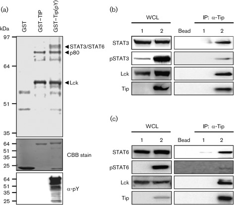
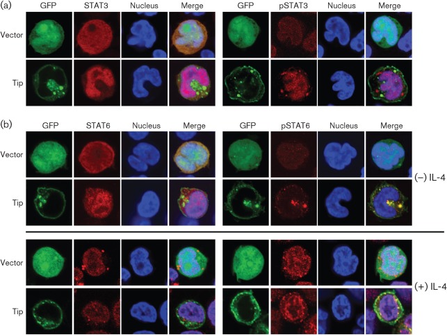
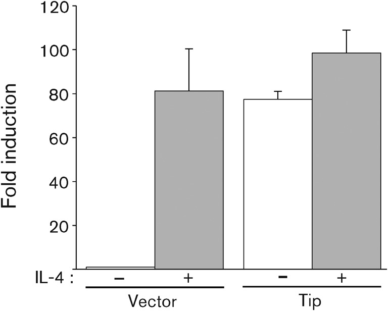
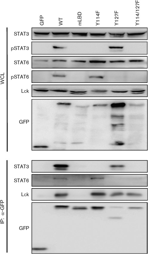
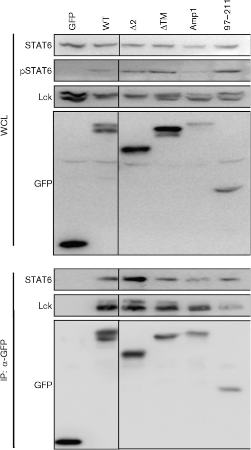
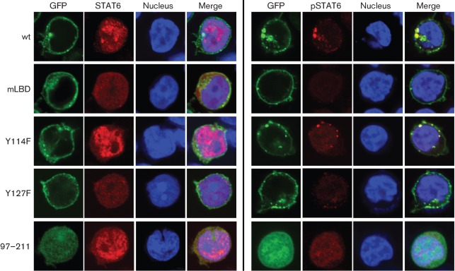
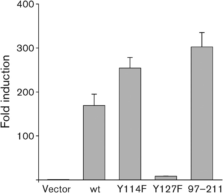
Herpesvirus saimiri (HVS), a T-lymphotropic monkey herpesvirus, induces fulminant T-cell lymphoma in non-natural primate hosts. In addition, it can immortalize human T-cells in vitro. HVS tyrosine kinase-interacting protein (Tip) is an essential viral gene required for T-cell transformation both in vitro and in vivo. In this study, we found that Tip interacts with the STAT6 transcription factor and induces phosphorylation of STAT6 in T-cells. The interaction with STAT6 requires the Tyr(127) residue and Lck-binding domain of Tip, which are indispensable for interleukin (IL)-2-independent T-cell transformation by HVS. It was also demonstrated that Tip induces nuclear translocation of STAT6, as well as activation of STAT6-dependent transcription in Jurkat T-cells. Interestingly, the phosphorylated STAT6 mainly colocalized with vesicles containing Tip within T-cells, but was barely detectable in the nucleus. However, nuclear translocation of phospho-STAT6 and transcriptional activation of STAT6 by IL-4 stimulation were not affected significantly in T-cells expressing Tip. Collectively, these findings suggest that the constitutive activation of STAT6 by Tip in T-cells may contribute to IL-2-independent T-cell transformation by HVS.


Figures

Fig. 1.
Fig. 1.
Tip interacts with STAT6. (a) To identify the proteins binding to Tip, a GST pull-down assay was performed with GST-fused phosphorylated Tip purified from E. coli strain TKX1 and Jurkat T-cell lysate. Expression of GST-tagged proteins and formation of phosphorylated protein were visualized by Coomassie brilliant blue (CBB) staining and immunoblotting using an anti-phosphotyrosine antibody (α-pY), respectively. Binding proteins were analysed by mass spectrometry. To confirm the interaction between Tip and STAT proteins, HEK293T cells were transfected with Lck and (b) STAT3 or (c) STAT6, together with (lane 2) or without (lane 1) Tip. Lysates were immunoprecipitated using anti-Tip antibody and immunocomplexes were analysed by immunoblotting using the indicated antibodies. Protein A/G beads without antibody were used to exclude non-specific binding (Bead). IP, Immunoprecipitation; WCL, whole-cell lysate.
Fig. 2.
Fig. 2.
Tip induces nuclear translocation of STAT6. Jurkat T-cells were electroporated with plasmids encoding GFP (vector) or GFP–Tip together with pVR/STAT3 or STAT6. Twenty-four hours after electroporation, localization of STAT3 (a) and STAT6 (b) was examined by confocal microscopy after staining with anti-STAT or anti-phosphoSTAT antibodes (red). TO-PRO-3 staining was used to visualize the nucleus (blue). Nuclear translocation of STAT6 was also examined in the absence (−) or presence (+) of IL-4 treatment (100 ng ml−1, 15 min).
Fig. 3.
Fig. 3.
Tip induces transcriptional activation of STAT6. Jurkat T-cells were electroporated with plasmids encoding Tip, STAT6 and 3×STAT6-luc plasmid and cultured in the presence (grey) or absence (white) of IL-4 (20 ng ml−1) for 18 h. To normalize transfection efficiency, pGK-βgal vector was included in the transfection mixture and fold induction of luciferase activity was determined after normalization with β-galactosidase activity. Each bar represents data from triplicate assays; error bars indicate sd.
Fig. 4.
Fig. 4.
The Tyr127 residue of Tip is required for the interaction with and phosphorylation of STAT6. Jurkat T-cells were electroporated with GFP, GFP–Tip or GFP–Tip mutant expression constructs. Twenty-four hours after electroporation, cells were lysed and used for immunoprecipitation (IP) with an antibody against GFP, followed by immunoblotting with the indicated antibodies. Whole-cell lysates (WCL) were used to detect the expression level of STATs and Lck.
Fig. 5.
Fig. 5.
Association of Tip with membrane lipid rafts is not required for its interaction with and phosphorylation of STAT6. Jurkat T-cells were electroporated with plasmids encoding Tip or its mutants that are defective in lipid-raft association or membrane trafficking. Twenty-four hours after electroporation, cells were lysed and used for immunoprecipitation (IP) with an antibody against GFP. Whole-cell lysates (WCL) and immunocomplexes were subjected to immunoblot assay using the indicated antibodies.
Fig. 6.
Fig. 6.
The Tyr127 residue of Tip is required for nuclear translocation of STAT6 by Tip. Jurkat T-cells were electroporated with plasmids encoding GFP-fused Tip wt or its mutants (green) together with pVR/STAT6. Twenty-four hours after electroporation, localization of STAT6 (left panels) and phosphoSTAT6 (right panels) was visualized by confocal microscopy after staining with anti-STAT6 or anti-phosphoSTAT6 antibodies (red). TO-PRO-3 staining was used to visualize the nucleus (blue).
Fig. 7.
Fig. 7.
The Tyr127 residue of Tip is required for transcriptional activation of STAT6. Jurkat T-cells were electroporated with plasmids encoding Tip wt or its mutants together with pVR/STAT6 and 3×STAT6-luc plasmid. Twenty-four hours after electroporation, luciferase activities were measured. To normalize transfection efficiency, the pGK-βgal vector was included in the transfection mixture and fold induction of luciferase activity was determined after normalization with β-galactosidase activity. Each bar represents data from triplicate assays; error bars indicate sd.

Similar articles

Cited by

References

    1. Benekli M., Baer M. R., Baumann H., Wetzler M. (2003). Signal transducer and activator of transcription proteins in leukemias. Blood 101, 2940–2954 10.1182/blood-2002-04-1204 - DOI - PubMed
    1. Biesinger B., Müller-Fleckenstein I., Simmer B., Lang G., Wittmann S., Platzer E., Desrosiers R. C., Fleckenstein B. (1992). Stable growth transformation of human T lymphocytes by herpesvirus saimiri. Proc Natl Acad Sci U S A 89, 3116–3119 10.1073/pnas.89.7.3116 - DOI - PMC - PubMed
    1. Biesinger B., Tsygankov A. Y., Fickenscher H., Emmrich F., Fleckenstein B., Bolen J. B., Bröker B. M. (1995). The product of the Herpesvirus saimiri open reading frame 1 (Tip) interacts with T cell-specific kinase p56lck in transformed cells. J Biol Chem 270, 4729–4734 10.1074/jbc.270.9.4729 - DOI - PubMed
    1. Bild A. H., Turkson J., Jove R. (2002). Cytoplasmic transport of Stat3 by receptor-mediated endocytosis. EMBO J 21, 3255–3263 10.1093/emboj/cdf351 - DOI - PMC - PubMed
    1. Bowman T., Garcia R., Turkson J., Jove R. (2000). STATs in oncogenesis. Oncogene 19, 2474–2488 10.1038/sj.onc.1203527 - DOI - PubMed

Publication types

MeSH terms

Substances