Comparative Study
. 2012 Jul;41(7):787-801.
doi: 10.1007/s00256-011-1284-2. Epub 2011 Oct 20.

Correlation between radiological assessment of acute ankle fractures and syndesmotic injury on MRI

Affiliations
Comparative Study

Correlation between radiological assessment of acute ankle fractures and syndesmotic injury on MRI

J J Hermans et al. Skeletal Radiol. 2012 Jul.

Abstract

Objective: Owing to the shortcomings of clinical examination and radiographs, injury to the syndesmotic ligaments is often misdiagnosed. When there is no indication requiring that the fractured ankle be operated on, the syndesmosis is not tested intra-operatively, and rupture of this ligamentous complex may be missed. Subsequently the patient is not treated properly leading to chronic complaints such as instability, pain, and swelling. We evaluated three fracture classification methods and radiographic measurements with respect to syndesmotic injury.

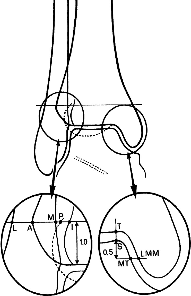
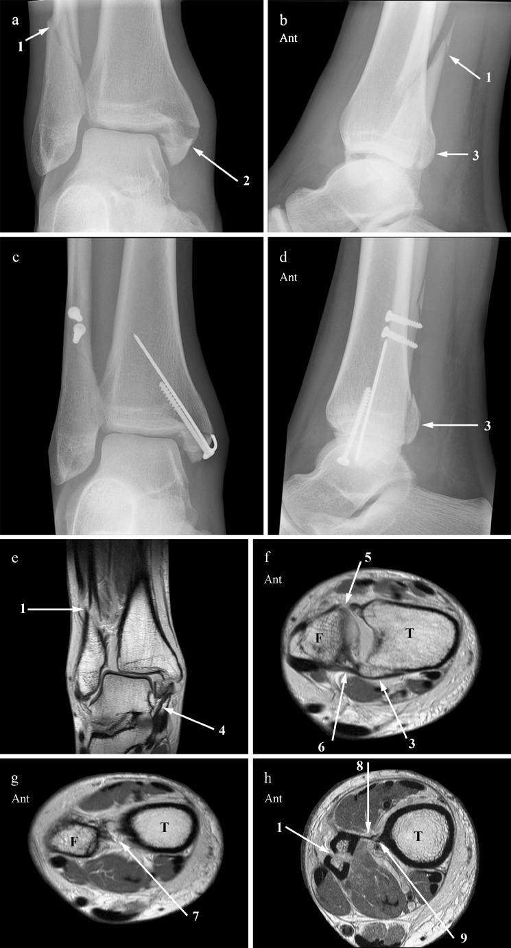
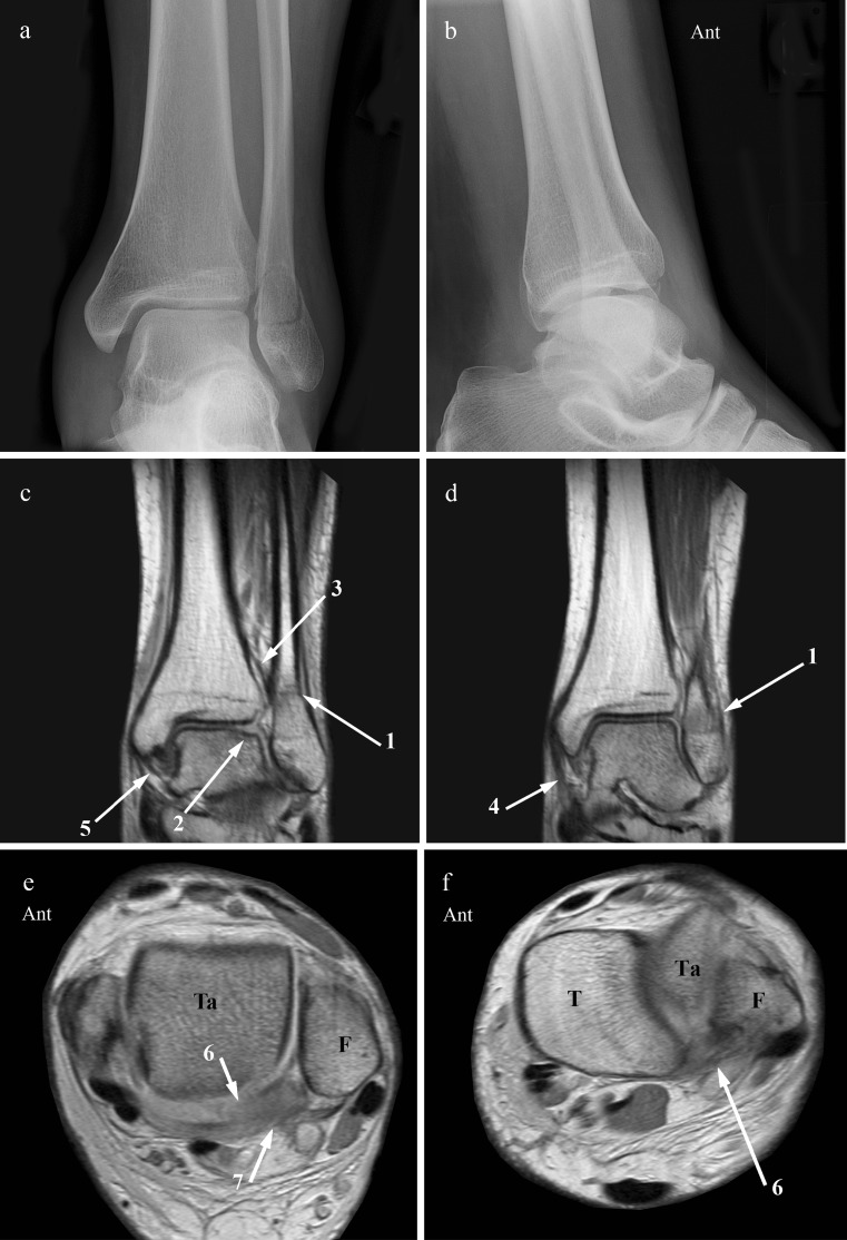
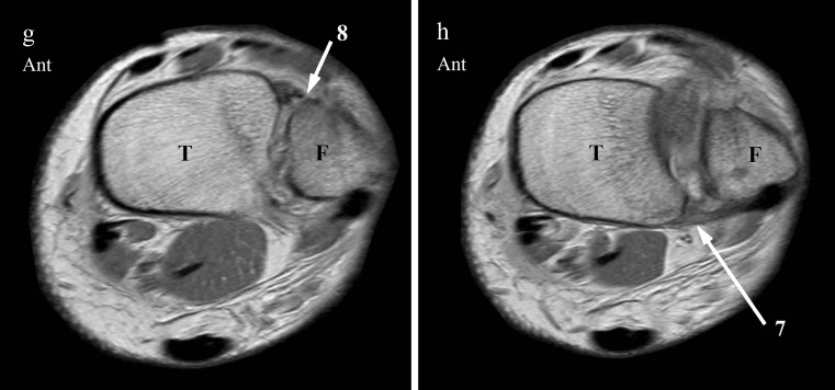
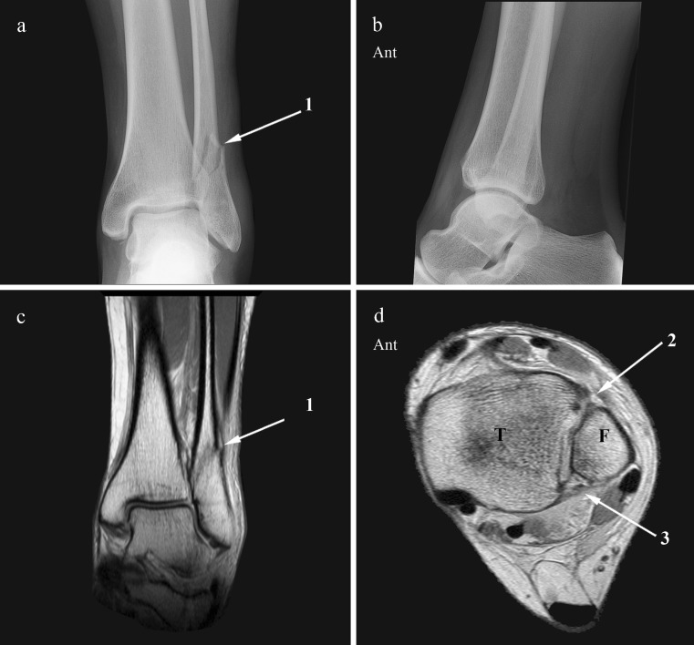
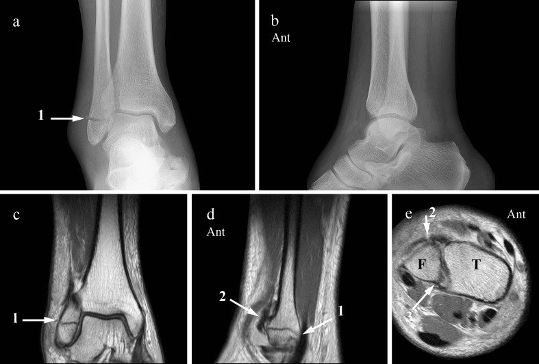
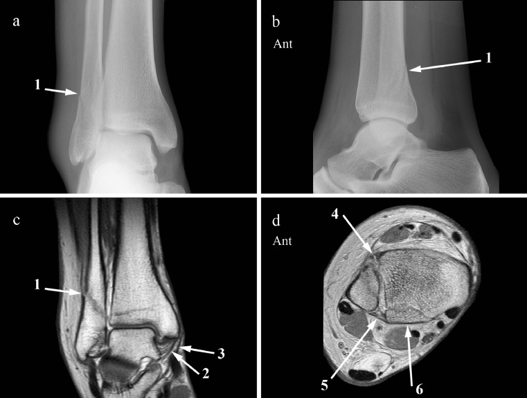
Materials and methods: Prospectively the radiographs of 51 consecutive ankle fractures were classified according to Weber, AO-Müller, and Lauge-Hansen. Both the fracture type and additional measurements of the tibiofibular clear space (TFCS), tibiofibular overlap (TFO), medial clear space (MCS), and superior clear space (SCS) were used to assess syndesmotic injury. MRI, as standard of reference, was performed to evaluate the integrity of the distal tibiofibular syndesmosis. The sensitivity and specificity for detection of syndesmotic injury with radiography were compared to MRI.

Results: The Weber and AO-Müller fracture classification system, in combination with additional measurements, detected syndesmotic injury with a sensitivity of 47% and a specificity of 100%, and Lauge-Hansen with both a sensitivity and a specificity of 92%. TFCS and TFO did not correlate with syndesmotic injury, and a widened MCS did not correlate with deltoid ligament injury.

Conclusion: Syndesmotic injury as predicted by the Lauge-Hansen fracture classification correlated well with MRI findings. With MRI the extent of syndesmotic injury and therefore fracture stage can be assessed more accurately compared to radiographs.


Figures

Fig. 1
Fig. 1
A schematic drawing of the ankle shows landmarks used for measurements of the different radiologic parameters. L Lateral border of the fibula, M medial border of the fibula, A anterior tibial tubercle, P posterior tibial tubercle, I floor of incisura fibularis, T tibial plafond, S superior point of medial talus, MT medial side of talus, LMM lateral side medial malleolus. AM is tibiofibular overlap (TFO). MI is tibiofibular clear space (TFCS). TS is superior clear space (SCS) and MTLMM is medial clear space (MCS). (Used with permission from A. Beumer, Clin. Orthop. Rel. Res 2004;423:227–234)
Fig. 2
Fig. 2
AP (a) and lateral (b) radiographs show an oblique fibula fracture (1), running from anteroinferior to posterosuperior, characteristic of a supination-external rotation injury. The medial malleolus (2) and posterior malleolus (3) are avulsed. AP (c) and lateral (d) radiographs after open reduction and internal fixation without a setscrew. The fracture of the posterior malleolus (3) is visible and not fixated. The fracture was classified as Weber type C, AO-Müller C2.3, Lauge-Hansen SE4. Coronal (e), 45° oblique (f, g), and axial proton-density-weighted (h) MR image. The coronal MRI shows the laterally dislocated talus, with a distal fibula fracture (1) and an avulsion fracture of the medial malleolus (2) attached to a thickened but intact superficial deltoid ligament (4). In f, the ATIFL (5) is ruptured, whereas the PTIFL (6) is intact and attached to an avulsion fracture of the posterolateral malleolus (3). In g, the ruptured interosseous ligament (7) is visible. In h, the fibula fracture (1) runs proximal to the interosseous membrane (8), which has a small tibial avulsion (9) up to this level. Ant Anterior, T tibia, F fibula
Fig. 3
Fig. 3
AP (a) and lateral (b) radiographs. Short distal fibula fracture (1) extending from just below to just above the level of the tibiotalar joint line. Measurements are normal. This fracture is classified as Weber type B, AO-Müller type B1.1, and Lauge-Hansen SE2. Coronal (c, d), axial (e) and 45° oblique (f, g, h) proton-density-weighted MR images. The coronal MRI (c, d) shows the fibula fracture (1), a posterolateral osteochondral lesion of the talar dome (2), a normal interosseous ligament (3), a thickened superficial (4), and a normal deep (5) deltoid ligament. On the axial (e) and oblique MR image (f), the transverse ligament (6) is ruptured. The PTIFL (7) appears to be ruptured in the axial plane (e) but is still continuous, although thickened, in the 45° oblique plane (h). The ATIFL (8) is ruptured (g). With the MRI findings, this would change the fracture into an AO-Müller type B1.2, but it would still be Lauge-Hansen SE2. Ant Anterior, Ta talus, T tibia, F fibula
Fig. 3
Fig. 3
AP (a) and lateral (b) radiographs. Short distal fibula fracture (1) extending from just below to just above the level of the tibiotalar joint line. Measurements are normal. This fracture is classified as Weber type B, AO-Müller type B1.1, and Lauge-Hansen SE2. Coronal (c, d), axial (e) and 45° oblique (f, g, h) proton-density-weighted MR images. The coronal MRI (c, d) shows the fibula fracture (1), a posterolateral osteochondral lesion of the talar dome (2), a normal interosseous ligament (3), a thickened superficial (4), and a normal deep (5) deltoid ligament. On the axial (e) and oblique MR image (f), the transverse ligament (6) is ruptured. The PTIFL (7) appears to be ruptured in the axial plane (e) but is still continuous, although thickened, in the 45° oblique plane (h). The ATIFL (8) is ruptured (g). With the MRI findings, this would change the fracture into an AO-Müller type B1.2, but it would still be Lauge-Hansen SE2. Ant Anterior, Ta talus, T tibia, F fibula
Fig. 4
Fig. 4
AP (a) and lateral (b) radiographs show a distal fibula fracture (1). Measurements are normal. The coronal proton-density-weighted MR image (c) also shows the fibula fracture (1). The axial proton-density-weighted MR image (d) is just below the level of the fibula fracture and demonstrates the rupture of the ATIFL (2). The fascicles of the PTIFL (3) are a little thickened but intact. This is a Weber type B, AO-Müller type B1.1, Lauge-Hansen SE2 fracture with normal measurements but with anterior syndesmotic injury. Ant Anterior, T tibia, F fibula
Fig. 5
Fig. 5
AP (a) and lateral (b) radiographs. A transverse fibular malleolar fracture (1) below the level of the tibiotalar joint space is visible. No fracture is visible at the medial or posterior malleolus. Measurements are normal. The fracture was classified as Weber A, AO-Müller A1.3, Lauge-Hansen SA1. Coronal (c), sagittal (d), and axial (e) proton-density-weighted MR image. The transverse fibula fracture (1) is visible on the coronal and sagittal MR image. The lower border of the ruptured ATIFL (2) lies just across the fibula fracture as can be seen on the sagittal MR image (d). In the 45° oblique image (e) the ATIFL (2) is thickened and avulsed from the fibula. The PTIFL (3) is intact. Ant Anterior, T tibia, F fibula
Fig. 6
Fig. 6
AP (a) and lateral (b) radiographs show a distal fibula fracture (1) at the level of the syndesmosis, running obliquely from anteroinferior to posterosuperior, characteristic of a supination-external rotation trauma. Measurements are normal. This fracture is classified as Weber type B, AO-Müller type B1.1, and Lauge-Hansen SE2. Coronal (c) and 45° oblique (d) proton-density-weighted MR images demonstrate the fibula fracture (1), and a normal deep (2) and superficial (3) deltoid ligament. The ATIFL is ruptured (4), whereas the intact PTIFL (5) is attached to an avulsion fracture of the posterolateral malleolus (6). This is therefore a Weber type B fracture with normal measurements but with anterior as well as posterior syndesmotic injury. According to Lauge-Hansen, this is an SE3 fracture

References

    1. Bartonicek J. Anatomy of the tibiofibular syndesmosis and its clinical relevance. Surg Radiol Anat. 2003;25(5–6):379–386. doi: 10.1007/s00276-003-0156-4. - DOI - PubMed
    1. Kelikian H, Kelikian S. Disorders of the ankle. London: W.B. Saunders Company; 1985.
    1. Kapandji IA. Funktionelle Anatomie der Gelenke. Schematisierte und kommentierte Zeichnungen zur menslichen Biomechanik. Stuttgart: Ferdinand Enke; 1985. 148–65.
    1. Lutz W. Zur Struktur der unteren Tibiofibularverbindung und der Membrana interossea cruris. Anat Entwickl Gesch. 1942;111:315–321. doi: 10.1007/BF00538090. - DOI
    1. Weber BG. Die Verletzungen des oberen Sprunggelenkes. Zweite, überarbeitete und ergänzte Auflage. Vienna: Hans Huber Bern Stuttgart; 1972.

Publication types