

Liposomes surface conjugated with human hemoglobin target delivery to macrophages
- PMID: 22012493
- PMCID: PMC3263929
- DOI: 10.1002/bit.24340
Liposomes surface conjugated with human hemoglobin target delivery to macrophages
Abstract
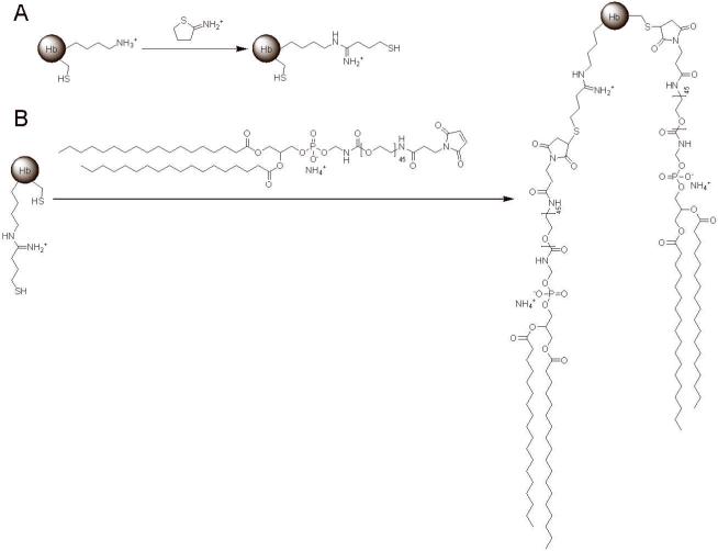
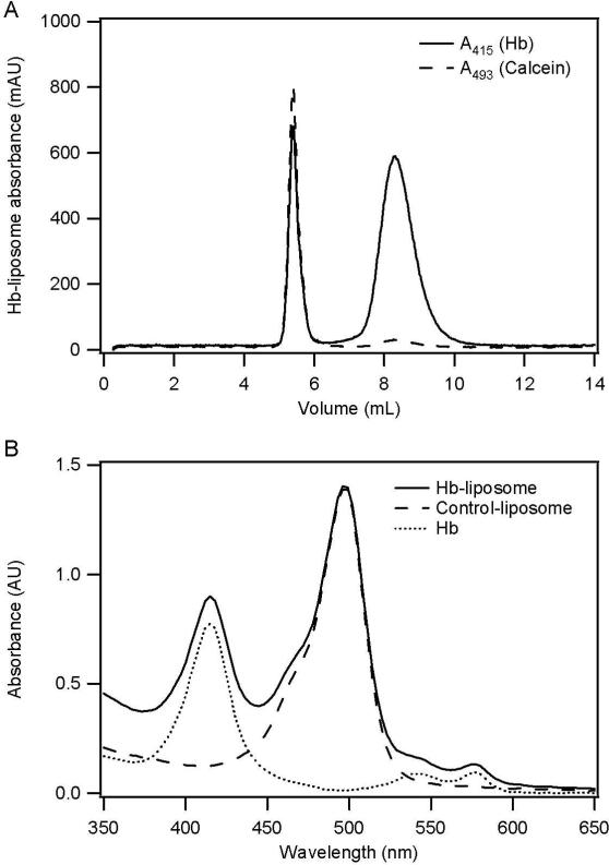
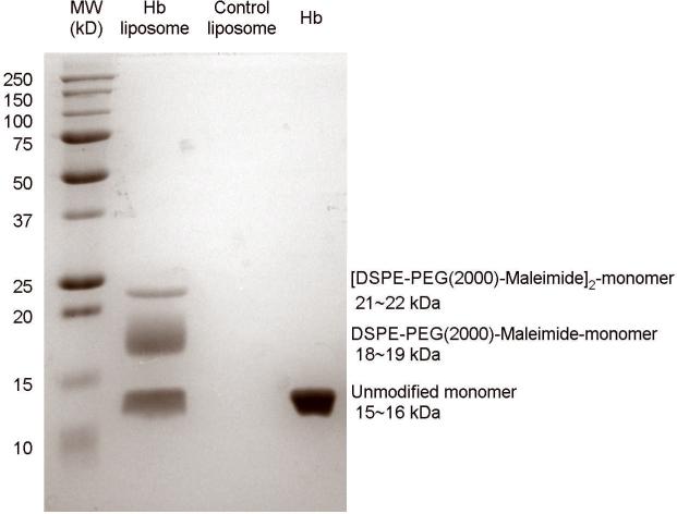
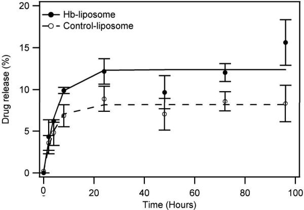
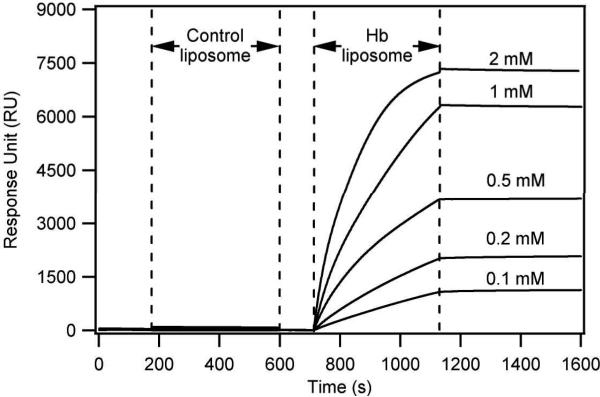
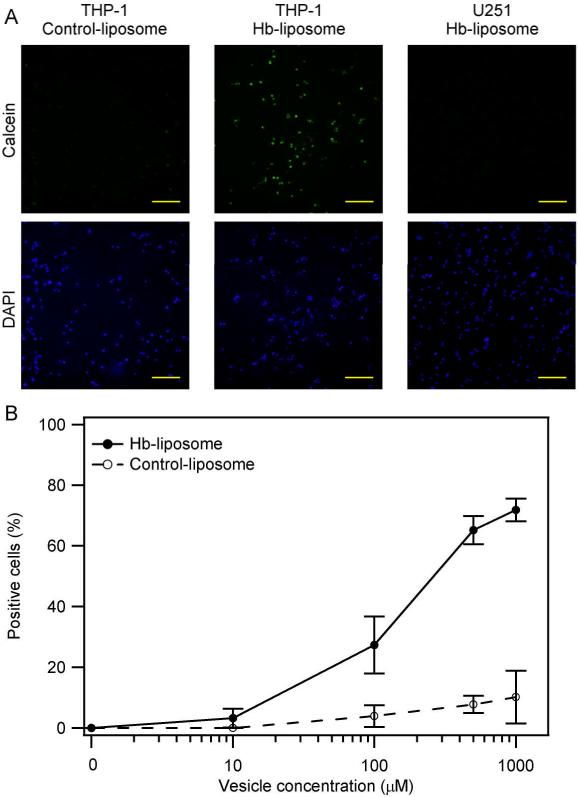
Current strategies to deliver therapeutic molecules to specific cell and tissue types rely on conjugation of antibodies and other targeting ligands directly to the therapeutic molecule itself or its carrier. This work describes a novel strategy to deliver therapeutic molecules into macrophages that takes advantage of the native hemoglobin (Hb) scavenging activity of plasma haptoglobin (Hp) and the subsequent uptake of the Hb-Hp complex into macrophages via CD163 receptor-mediated endocytosis. The drug delivery system described in this work consists of Hb decorated liposomes that can encapsulate any therapeutic molecule of interest, in this case the model fluorescent dye calcein was used in this study. The results of this study clearly demonstrate that this delivery system is specific towards macrophages and demonstrates the feasibility of using this approach in targeted drug delivery.
Copyright © 2011 Wiley Periodicals, Inc.
Figures

Similar articles
-
Efficient intracellular drug-targeting of macrophages using stealth liposomes directed to the hemoglobin scavenger receptor CD163.J Control Release. 2012 May 30;160(1):72-80. doi: 10.1016/j.jconrel.2012.01.034. Epub 2012 Jan 27. J Control Release. 2012. PMID: 22306335
-
Hyperglycemia Induces Inflammatory Response of Human Macrophages to CD163-Mediated Scavenging of Hemoglobin-Haptoglobin Complexes.Int J Mol Sci. 2022 Jan 26;23(3):1385. doi: 10.3390/ijms23031385. Int J Mol Sci. 2022. PMID: 35163309 Free PMC article.
-
Soluble macrophage-derived CD163: a homogenous ectodomain protein with a dissociable haptoglobin-hemoglobin binding.Immunobiology. 2010 May;215(5):406-12. doi: 10.1016/j.imbio.2009.05.003. Epub 2009 Jul 5. Immunobiology. 2010. PMID: 19581020
-
CD163: a signal receptor scavenging haptoglobin-hemoglobin complexes from plasma.Int J Biochem Cell Biol. 2002 Apr;34(4):309-14. doi: 10.1016/s1357-2725(01)00144-3. Int J Biochem Cell Biol. 2002. PMID: 11854028 Review.
-
The haptoglobin-CD163-heme oxygenase-1 pathway for hemoglobin scavenging.Oxid Med Cell Longev. 2013;2013:523652. doi: 10.1155/2013/523652. Epub 2013 May 27. Oxid Med Cell Longev. 2013. PMID: 23781295 Free PMC article. Review.
Cited by
-
The Role of Tumor Epithelial-Mesenchymal Transition and Macrophage Crosstalk in Cancer Progression.Curr Osteoporos Rep. 2023 Apr;21(2):117-127. doi: 10.1007/s11914-023-00780-z. Epub 2023 Feb 27. Curr Osteoporos Rep. 2023. PMID: 36848026 Free PMC article. Review.
-
Tumor-Associated Macrophage Targeting of Nanomedicines in Cancer Therapy.Pharmaceutics. 2023 Dec 29;16(1):61. doi: 10.3390/pharmaceutics16010061. Pharmaceutics. 2023. PMID: 38258072 Free PMC article. Review.
-
Harnessing molecular recognition for localized drug delivery.Adv Drug Deliv Rev. 2021 Mar;170:238-260. doi: 10.1016/j.addr.2021.01.008. Epub 2021 Jan 20. Adv Drug Deliv Rev. 2021. PMID: 33484737 Free PMC article. Review.
-
Macrophages associated with tumors as potential targets and therapeutic intermediates.Nanomedicine (Lond). 2014 Apr;9(5):695-707. doi: 10.2217/nnm.14.13. Nanomedicine (Lond). 2014. PMID: 24827844 Free PMC article. Review.
-
Full-length in meso structure and mechanism of rat kynurenine 3-monooxygenase inhibition.Commun Biol. 2021 Feb 4;4(1):159. doi: 10.1038/s42003-021-01666-5. Commun Biol. 2021. PMID: 33542467 Free PMC article.
References
-
- Brookes S, Biessels P, Ng NF, Woods C, Bell DN, Adamson G. Synthesis and characterization of a hemoglobin-ribavirin conjugate for targeted drug delivery. Bioconjug Chem. 2006;17(2):530–7. - PubMed
-
- Cao Z, Tong R, Mishra A, Xu W, Wong GC, Cheng J, Lu Y. Reversible cell-specific drug delivery with aptamer-functionalized liposomes. Angew Chem Int Ed Engl. 2009;48(35):6494–8. - PubMed
-
- Danoff EJ, Wang X, Tung SH, Sinkov NA, Kemme AM, Raghavan SR, English DS. Surfactant vesicles for high-efficiency capture and separation of charged organic solutes. Langmuir. 2007;23(17):8965–71. - PubMed
-
- Elmer J, Buehler PW, Jia YP, Wood F, Harris DR, Alayash AI, Palmer AF. Functional Comparison of Hemoglobin Purified by Different Methods and Their Biophysical Implications. Biotechnology and Bioengineering. 2010;106(1):76–85. - PubMed
Publication types
MeSH terms
Substances
Grants and funding
LinkOut - more resources
Full Text Sources
Other Literature Sources
Research Materials
Miscellaneous
