. 2011 Oct 15;25(20):2158-72.
doi: 10.1101/gad.16962311.

The Cyclin K/Cdk12 complex maintains genomic stability via regulation of expression of DNA damage response genes

Affiliations

The Cyclin K/Cdk12 complex maintains genomic stability via regulation of expression of DNA damage response genes

Dalibor Blazek et al. Genes Dev. .

Abstract

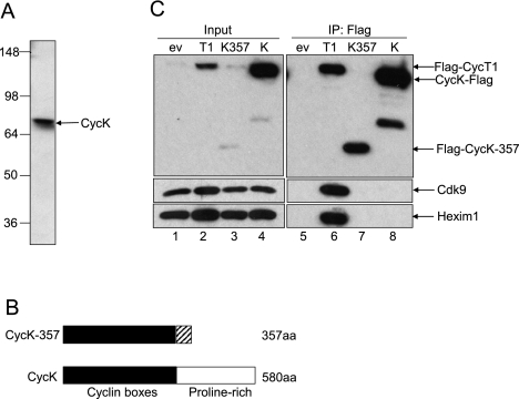
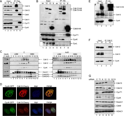
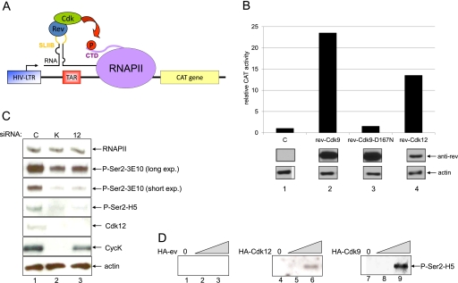
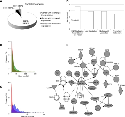
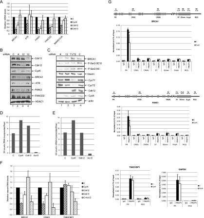
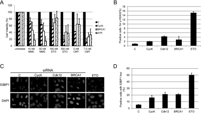
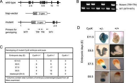
Various cyclin-dependent kinase (Cdk) complexes have been implicated in the regulation of transcription. In this study, we identified a 70-kDa Cyclin K (CycK) that binds Cdk12 and Cdk13 to form two different complexes (CycK/Cdk12 or CycK/Cdk13) in human cells. The CycK/Cdk12 complex regulates phosphorylation of Ser2 in the C-terminal domain of RNA polymerase II and expression of a small subset of human genes, as revealed in expression microarrays. Depletion of CycK/Cdk12 results in decreased expression of predominantly long genes with high numbers of exons. The most prominent group of down-regulated genes are the DNA damage response genes, including the critical regulators of genomic stability: BRCA1 (breast and ovarian cancer type 1 susceptibility protein 1), ATR (ataxia telangiectasia and Rad3-related), FANCI, and FANCD2. We show that CycK/Cdk12, rather than CycK/Cdk13, is necessary for their expression. Nuclear run-on assays and chromatin immunoprecipitations with RNA polymerase II on the BRCA1 and FANCI genes suggest a transcriptional defect in the absence of CycK/Cdk12. Consistent with these findings, cells without CycK/Cdk12 induce spontaneous DNA damage and are sensitive to a variety of DNA damage agents. We conclude that through regulation of expression of DNA damage response genes, CycK/Cdk12 protects cells from genomic instability. The essential role of CycK for organisms in vivo is further supported by the result that genetic inactivation of CycK in mice causes early embryonic lethality.


Figures

Figure 1.
Figure 1.
Identification of CycK as 70-kDa protein in cells. (A) Detection of the 70-kDa form of CycK in cell lysates from 293 cells with an antibody directed against the N-terminal cyclin box of CycK (Sigma, HPA000645). (B) Schematic representation of 40-kDa CycK-357 (Edwards et al. 1998) and the 70-kDa forms of CycK. (C) CycK and CycK-357 do not bind Cdk9. Flag-CycT1, CycK-Flag, Flag-CycK-357, and Flag-ev were immunoprecipitated from 293 cells and probed with antibodies to Cdk9 (right middle panel) and Hexim1 (right bottom panel) by Western blotting. (Left panels) Expression of the Cdk9, Hexim1, and Flag epitope-tagged proteins was measured with appropriate antibodies and represents 5% input of cell lysates.
Figure 2.
Figure 2.
CycK binds Cdk12 and Cdk13 in two separate complexes. (A) CycK interacts with Cdk12 and Cdk13, but not with Cdk9. (Lanes 4–6) Flag epitope-tagged proteins or empty plasmid vector (ev) were immunoprecipitated from lysates of 293 cells, immunoprecipitations were resolved by Western blotting, and bound proteins were identified with antibodies indicated on the right. Lanes 1–3 represent 5% input of cell lysates. (B) Cdk12 and Cdk13 interact with CycK, rather than with CycL. (Lanes 5–8) HA epitope-tagged proteins or empty plasmid vector (ev) were immunoprecipitated from lysates of 293 cells, immunoprecipitations were resolved by SDS-PAGE, and bound proteins were identified with antibodies indicated on the right by Western blotting. Lanes 1–4 represent 5% input of the cell lysates to the immunoprecipitation. Stars indicate the positions of HA epitope-tagged proteins. (C) CycK, Cdk12, and Cdk13 comigrate in glycerol gradient. Lysates of 293 cells from control (left panel) or actinomycin D-treated (right panel) cells were divided into 13 fractions by a glycerol gradient centrifugation. Amounts of endogenous proteins were assessed with antibodies indicated on the inner sides of both panels by Western blotting. Numbers above and below the panels refer to the glycerol gradient fraction. (LMM) Low molecular mass; (HMM) high molecular mass; (ActD) actinomycin D; (C) control. Five percent input of lysates in glycerol gradients is presented in the panel on the left. (D) CycK colocalizes with Cdk12 and Cdk13. Expression of GFP- and Cherry-tagged proteins in HeLa cells was visualized by confocal microscopy either alone or merged as indicated above the pictures. DAPI depicts for the staining of the nucleus. (E) The CycK/Cdk13 complex is free of Cdk12. (Lanes 3,4) Cdk13-HA or HA-ev were immunoprecipitated from the lysate of 293 cells and immunoprecipitations were resolved by SDS-PAGE followed by Western blotting, where bound proteins were identified with antibodies indicated on the right. Lanes 1 and 2 represent 5% input of cell lysate. (F) The CycK/Cdk12 complex is free of Cdk13. Endogenous Cdk12 or control (C) immunoprecipitation without antibody was carried out as in E. (Lanes 3,4) Proteins identified by Western blotting are indicated on the right. Lanes 1 and 2 represent 5% input of cell lysate. (G) CycK stabilizes CycK/Cdk12 and CycK/Cdk13 complexes. Proteins were knocked down in HCT116 cells by the indicated siRNAs and lysates were separated by SDS-PAGE followed by Western blotting with the antibodies indicated on the side of the panels.
Figure 3.
Figure 3.
CycK/Cdk12 regulates transcription and phosphorylates Ser2 in the CTD of RNAPII. (A) Schematic depiction of heterologous RNA tethering assay. Plasmid reporter used to test transcription contains modified HIV-LTR promoter (blue square) followed by TAR (transactivation response RNA) (red square) with inserted stem–loop IIB (SLIIB) (yellow line in RNA) from the HIV Rev response element (RRE). Nascent RNA (black dashed line) synthetized by RNAPII (violet oval) forms a dsRNA loop with the SLIIB element. Rev-cdk fusion proteins tethered via SLIIB to paused RNAPII can release RNAPII when phosphorylate Ser2 (P) (red circle) in its CTD (violet line), which results in the transcription of CAT reporter gene (yellow square). (B) Rev-Cdk12 activates transcription from the SLIIB-CAT plasmid. The activity of the CAT reporter SLIIB-CAT coexpressed with empty plasmid vector (C) was set as 1, and bars represent relative CAT activity obtained by the cotransfection of the reporter with indicated plasmids in HeLa cells. Western blotting shows expression of Rev fusion proteins and actin. (C) Knockdown of CycK/Cdk12 decreases the global phosphorylation of Ser2 in the CTD of RNAPII. HCT116 cell were transfected with the indicated siRNAs, and the levels of proteins were followed with the indicated antibodies by Western blotting. (D) CycK/Cdk12 phosphorylates Ser2 in the GST-CTD in vitro. Ten nanograms of GST-CTD was incubated with increasing amounts (indicated by the triangle) of purified HA-ev, Cdk12-HA, and Cdk9-HA. The amounts of phosphorylated Ser2 (P-Ser2) were monitored by Western blotting with the indicated anti-Ser2 phospho-specific antibody.
Figure 4.
Figure 4.
CycK/Cdk12 knockdown changes expression of a small subset of human genes. (A) Distribution of genes either differentially expressed or with no change of expression after CycK knockdown in HeLa cells. (B) The graph presents the distributions of the length of all human genes (red) and genes down-regulated in expression microarray in Cdk12-depleted cells (green). Human gene length data were obtained from the University of California at Santa Cruz (UCSC) Genome Browser. (C) The graph presents the distributions of the number of exons in all human genes (red) and genes down-regulated in expression microarray in Cdk12-depleted cells (blue). Data about the number of exons were obtained from the UCSC browser. (D) The classification of genes with reduced expression after CycK knockdown is based on their enrichment relative to total numbers in their specific category. Significance is expressed as −log (P-value) with a threshold value of 1.3 = −log (P = 0.05), and was calculated by the Ingenuity program using right-tailed Fischer's exact test. (E) Depiction of the BRCA1 network. The network was generated by the Ingenuity program from genes down-regulated after CycK knockdown. (Gray shapes) Genes down-regulated in the microarray; (white shapes) genes not found down-regulated in the microarray; (oval) transcriptional regulator; (diamond) enzyme; (triangle) kinase; (trapezoid) transporter. A solid line represents direct interaction, a dashed line indicates indirect interaction, a line without arrows indicates binding, and an arrow from protein A to protein B means that protein A acts on protein B.
Figure 5.
Figure 5.
CycK/Cdk12 regulates transcription of key DDR genes. (A) Knockdown of CycK/Cdk12 results in decreased mRNA levels of key DDR genes in HCT116 cells. Graphs present levels of mRNA of described genes in cells transfected with control (C), CycK, Cdk12, or Cdk13 siRNA. RT-qPCR results are normalized to the mRNA of GAPDH. (B) Knockdown of CycK/Cdk12, rather than of CycK/Cdk13, depletes protein levels of the DDR genes. HCT116 cell were transfected with indicated siRNAs, and protein levels were measured with indicated antibodies by Western blotting. (C) Knockdown of P-TEFb does not decrease expression of the BRCA1 gene. HCT116 cells were transfected with the depicted siRNAs, and protein levels were measured by Western blotting with the indicated antibodies. (D,E) Depletion of CycK/Cdk12 in cells does not affect the global transcription rate in nascent RNA analysis. HeLa cells were transfected with indicated siRNA for 48 h or treated with actinomycin D for 1 h. The graphs present the ratio of RNA-incorporated 3H-uridine in precipitated nascent RNA (D) or in polyadenylated nascent mRNA (E) to free 3H-uridine as a measure of the amount of cells. Representative experiments are shown. (F) Reduction of nascent transcript of BRCA1 and FANCI in nuclear run-on from CycK/Cdk12-depleted cells. HCT116 cells were transfected with indicated siRNA or treated with actinomycin D. Graphs show relative abundance of nascent RNA transcripts for indicated genes in nuclear run-on measured by RT-qPCR. RT-qPCR results are normalized to the nascent mRNA of GAPDH, and error bars indicate the standard deviation from three independent transfections. (G) Depletion of the CycK leads to lower levels of the RNAPII on the promoters of BRCA1 and FANCI genes. ChIP analysis for the occupation of RNAPII on the regions of indicated genes after transfection with mock or CycK siRNAs. Schemes depicting BRCA1 and FANCI genes show the position of ChIP primers. Their location is marked by the arrows, and the number indicates their approximate distance in kilobases from the transcription start site. (PR) Promotor; [CR(5)] 5′ prime of the coding region; [CR(M)] coding region; (ST) stop codon; (3Down) region down of the stop codon; (PolyA) polyadenylation signal; [IR(3)] intergenic region. IgG corresponds to the empty beads control. Experiments are the results of four (BRCA1 and FANCI) or three (TNSK1BP1 and GAPDH) independent transfections of HCT116 cells, and qPCR was performed in triplicate for each transfection. Statistical significance of each pairwise comparison is depicted with a star; (*) P < 0.05.
Figure 6.
Figure 6.
CycK/Cdk12 is required for the maintenance of genomic stability. (A) CycK-depleted cells are sensitive to a variety of DNA damage agents. The graphs represent the results of survival assays of HeLa cells transfected with siRNA directed to the shown targets and treated with either mitomycin C (MMC), etoposide (ETO), or camptothecin (CMT), or left untreated. Mean and standard deviation values represent the result of at least three independent transfections. Cell viability was normalized to relative growth of cells transfected with specific siRNA. (B) Depletion of CycK/Cdk12 induces DNA damage signaling. The graph shows percentage of positive cells × median fluorescence intensity of HeLa cells transfected with indicated siRNAs or treated with etoposide (ETO), labeled with γ-H2AX antibody, and analyzed by fluorescence-activated cell counting. The results represent the values of four independent experiments. (C,D) Knockdown of the CycK/Cdk12 complex causes spontaneous DNA damage. (C) U2OS cells transfected with the indicated siRNAs or treated with etoposide (ETO) were labeled with 53BP1 antibody or DAPI, and images showing 53BP1 foci were visualized by indirect immunofluorescence. (D) Graph represents the quantification of cells positive for 53BP1 foci from three independent experiments. Cells with at least five foci were considered positive, and a minimum of 300 cells were analyzed for each condition in each experiment.
Figure 7.
Figure 7.
Genetic inactivation of CycK leads to early embryonic lethality. (A) Schematic representation of wild-type (WT) and trapped (TR) (CycK.β-geo) alleles. Wild-type and trapped loci contain one to 11 and one to four exons, respectively. The integration of the gene trap vector into intron 1 of CycK resulted in the inactivation of the CycK gene. The insertion also led to an in-frame fusion of CycK with the β-geo (encoding β-galactosidase and neomycin phosphotransferase) reporter gene. Arrows below the scheme represent primer sets for wild-type (WT) (WTf and WTr) and trapped (TR) (TRf and TRr) alleles used in B. (B) Representative result of PCR genotyping. PCR genotyping of genomic DNA from the adult mouse was performed with two sets of primers—WTf+WTr and TRf+TRr (shown in A, below the mutant allele of the CycK gene)—to detect wild-type (lower band) and trapped (upper band) alleles of the CycK gene. Genomic DNA was isolated from 3-wk-old mice. For the primers used, see the Supplemental Material. (C) The table presents the results of genotyping of specified CycK embryos at indicated embryonic days. (D) Expression of CycK during mouse mid-gestation and early embryogenesis. LacZ staining of heterozygous embryos was used to visualize spatiotemporal expression of CycK.β-geo, which reflects transcription from the endogenous CycK promoter. Wild-type (+/+) and heterozygous (+/−) embryos were isolated at the given embryonic day and stained with X-Gal. CycK was expressed in forming tissues and organs in E8.5 and E11.5 embryos. Importantly, the expression of CycK was localized to the embryonic region and ectoderm in E6.5 and E7.5 embryos.

Comment in

Similar articles

Cited by

References

    1. Ahn SH, Kim M, Buratowski S 2004. Phosphorylation of serine 2 within the RNA polymerase II C-terminal domain couples transcription and 3′ end processing. Mol Cell 13: 67–76 - PubMed
    1. Bartkowiak B, Liu P, Phatnani HP, Fuda NJ, Cooper JJ, Price DH, Adelman K, Lis JT, Greenleaf AL 2010. CDK12 is a transcription elongation-associated CTD kinase, the metazoan ortholog of yeast Ctk1. Genes Dev 24: 2303–2316 - PMC - PubMed
    1. Bell D, Berchuck A, Birrer M, Chien J, Cramer DW, Dao F, Dhir R, DiSaia P, Gabra H, Glenn P, et al. 2011. Integrated genomic analyses of ovarian carcinoma. Nature 474: 609–615 - PMC - PubMed
    1. Blazek D, Barboric M, Kohoutek J, Oven I, Peterlin BM 2005. Oligomerization of HEXIM1 via 7SK snRNA and coiled-coil region directs the inhibition of P-TEFb. Nucleic Acids Res 33: 7000–7010 - PMC - PubMed
    1. Bonner WM, Redon CE, Dickey JS, Nakamura AJ, Sedelnikova OA, Solier S, Pommier Y 2008. γH2AX and cancer. Nat Rev Cancer 8: 957–967 - PMC - PubMed

Publication types