. 2011 Oct 15;25(20):2187-97.
doi: 10.1101/gad.16974811.

Prox1 dosage controls the number of lymphatic endothelial cell progenitors and the formation of the lymphovenous valves

Affiliations

Prox1 dosage controls the number of lymphatic endothelial cell progenitors and the formation of the lymphovenous valves

R Sathish Srinivasan et al. Genes Dev. .

Abstract

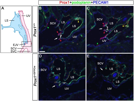
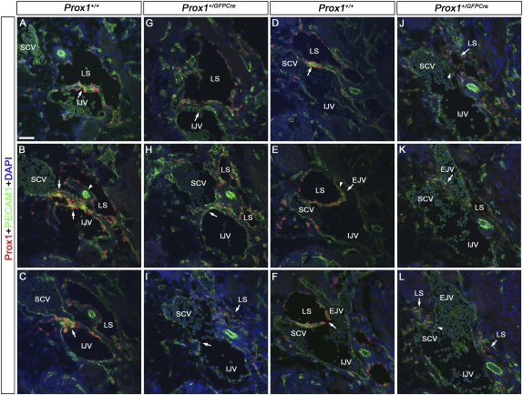
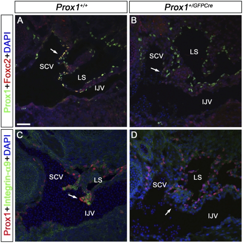
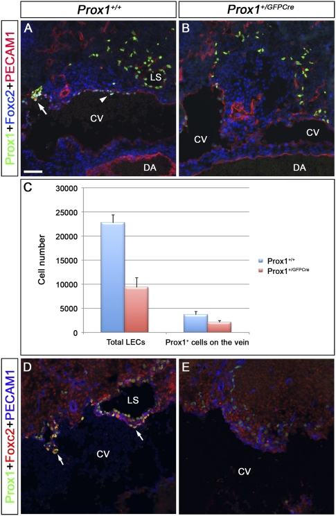
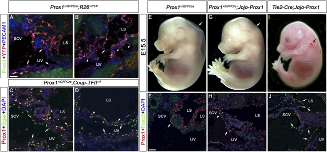
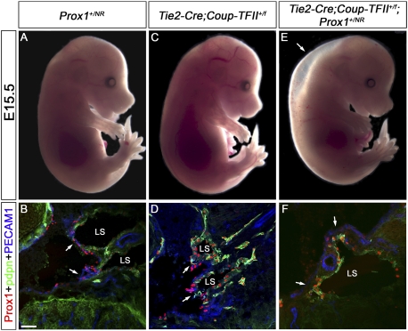
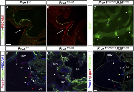
Arteries, veins, and lymphatic vessels are functionally linked, and their physical interaction is tightly regulated. The lymphatic vessels communicate with the blood vessels only at the junction of the jugular and subclavian veins. Here, we characterize the embryonic lymphovenous valves controlling this vital communication and show that they are formed by the intercalation of lymphatic endothelial cells (LECs) with a subpopulation of venous endothelial cells (ECs) at the junction of the jugular and subclavian veins. We found that unlike LEC progenitors, which move out from the veins and differentiate into mature LECs, these Prox1-expressing ECs remain in the veins and do not acquire LEC features. We demonstrate that the development of this Prox1-expressing venous EC population, and therefore of lymphovenous valves, requires two functional copies of Prox1, as the valves are absent in Prox1 heterozygous mice. We show that this is due to a defect in the maintenance of Prox1 expression in venous ECs and LEC progenitors promoted by a reduction in Coup-TFII/Prox1 complex formation. This is the first report describing the molecular mechanism controlling lymphovenous communication.


Figures

Figure 1.
Figure 1.
Prox1 is expressed on a previously unidentified population of ECs in the lymphovenous valves. (A) Schematic representation (adapted with modifications from van der Putte, 1975) of the area of an E13.5 embryo where the jugular and subclavian veins join to form the lymphovenous valves. The head (anterior) is oriented toward the top and the heart (posterior) is oriented toward the bottom of the figure. These veins merge to form the SVC. The posterior portion of the jugular lymph sac (LS) opens into the SCV where the veins merge. To visualize the boxed region of the diagram, E13.5 wild-type (B,C) or Prox1+/GFPCre (D,E) embryos were frontally sectioned from the dorsal to the ventral side and immunostained for the LEC markers Prox1 and podoplanin and the pan-endothelial PECAM1. (B) The first valve (arrow) is adjacent to the IJV on one side and the SCV on the other side. (C) The second valve (arrow) in this orientation is on the SCV. Each of the valve's two leaflets consists of two layers of Prox1+ ECs: an inner Prox1+ podoplanin+ layer that is continuous with the lymph sac (white arrowheads) and an outer Prox1+ podoplanin layer that is continuous with the veins (red arrowheads). Note the relatively high levels of Prox1 in the valves (red arrowheads) and in some cells of the lymph sac (yellow arrowheads). (B,C) Few Prox1+ cells are also seen on the veins away from the lymphovenous valves (green arrowheads). (D) In Prox1+/GFPCre embryos, Prox1 is not expressed on the walls of the veins (arrow). (E) Occasionally, the lymph sacs abruptly fuse with the IJV (arrow). The brain is oriented toward the right, the heart is oriented toward the left, and the thymus is oriented toward the bottom of B–E. Bar, 50 μm.
Figure 2.
Figure 2.
Lymphovenous valves are formed by the fusion of lymph sacs with two adjacent veins. E13.5 wild-type (A–F) or Prox1+/GFPCre (G–L) embryos were transversely sectioned in an anterior to posterior orientation in the region where the jugular and subclavian veins interact (box in Fig. 1A) and were immunostained for the LEC marker Prox1 and the pan-endothelial marker PECAM1. (A) In wild-type embryos, Prox1 is expressed uniformly in LECs forming the lymph sac (LS) and in a polarized manner on the IJV (arrow). Note the relatively high levels of Prox1 on the ECs in the vein and in the lymph sacs' LECs that are facing the vein. (B) The lymph sac is split into two portions by the vertebral artery (white arrowhead). Both walls of the medial portion of the lymph sac intercalate with the wall of the IJV medially and the SCV laterally (arrows). (C) The IJV and the SCV have completely merged together, and the valve rudiment is seen in the middle (arrow). (D) The lateral portion of the lymph sac (LS) runs adjacent to the SCV. Note the relatively higher levels of Prox1 in the venous ECs and in the LECs in the lymph sac facing the vein (arrow). (E) The EJV is branching off from the SCV (arrow), and Prox1 is expressed on the walls of this vein in a polarized manner (arrowhead). This wall is also adjacent to the lymph sac. (F) The opening of the valve (arrow) is now seen and is formed by the fusion of the two layers of Prox1+ ECs. (G,H) In Prox1+/GFPCre embryos, Prox1 is expressed in LECs forming the lymph sac, but very few Prox1+ cells are seen on the walls of the IJV and SCV (arrows). (I) At the point where the IJV and the SCV merge, no Prox1+ cells or valve-like structures exist (arrow). (J) No Prox1 expression was observed on the wall of the SCV that lies close to the lymph sac (arrowhead). (K) Posterior to that, the EJV branches off from the SCV. No Prox1+ cells are seen on the walls of the EJV (arrow). (L) No communication is observed between the lymph sac and the veins (arrowhead). Also, note an overall reduction in the number of LECs in G–L. The neural tube is oriented toward the right, the heart is oriented toward the left, and the thymus is oriented toward the bottom in each panel. Bar, 50 μm.
Figure 3.
Figure 3.
Foxc2 and Integrin-α9 are expressed in the lymphovenous valves but are absent in Prox1 heterozygous embryos. E13.5 wild-type (A,C) and Prox1+/GFPCre (B,D) embryos were frontally sectioned and immunostained for either Prox1 and Foxc2 (A,B) or Prox1 and Integrin-α9 (C,D). (A) In wild-type embryos, Foxc2 is specifically expressed on the outer layer of the valve's leaflets, which are continuous with the walls of the veins (arrow). (B) In sections from an identical region in Prox1+/GFPCre embryos, Foxc2 is not expressed on the walls of the veins (arrow). Integrin-α9 is strongly expressed in the valves of control embryos (C, arrow) but is absent in Prox1+/GFPCre littermates (D, arrow). The head is oriented toward the right, the heart is oriented toward the left, and the thymus is oriented toward the bottom in both panels. (IJV) Internal jugular vein; (SCV) subclavian vein; (LS) lymph sac. Bar, 50 μm.
Figure 4.
Figure 4.
The number of Prox1-expressing LEC progenitors and Prox1-expressing venous ECs that will form the lymphovenous valves is reduced in Prox1 heterozygous embryos. (A,B) E11.5 control (A) and Prox1+/GFPCre (B) embryos were frontally sectioned and immunostained for Prox1, Foxc2, and PECAM1. (A) In the control embryos, Foxc2 is weakly expressed in the Prox1+ cells on the vein (arrowhead). In addition, lymph sacs and occasionally one valve rudiment (arrow) could be observed. (B) In Prox1+/GFPCre embryos, the total number of LECs appears to be reduced, and no lymph sacs or valve rudiments are seen. (C) Compared with wild-type littermates, the total number of Prox1+ PECAM1+ ECs on the vein and outside the vein were reduced in E11.5 Prox1+/GFPCre embryos (n = 3 for each genotype, P < 0.05). (D,E) E12.5 control (D) and Prox1+/GFPCre (E) embryos were frontally sectioned and immunostained for Prox1, Foxc2, and PECAM1. (D) In control embryos, Foxc2 is expressed on the valve rudiments (arrows), which lie adjacent to the primitive lymph sacs. (E) In Prox1+/GFPCre embryos, the total number of LECs appears to be reduced, and no valve rudiments are seen. Lymph sacs are also not observed in this location. The head is oriented toward the right, the heart is oriented toward the left, and the thymus is oriented toward the bottom in all panels. (CV) Cardinal vein; (DA) dorsal aorta; (LS) lymph sac. Bar, 50 μm.
Figure 5.
Figure 5.
Prox1 heterozygous embryos do not maintain Prox1 expression in a subpopulation of LEC and lymphovenous valve progenitors. (A,B) E13.5 Prox1+/GFPCre;R26+/YFP embryos were sectioned and immunostained for Prox1, YFP, and PECAM1. (A) No Prox1+ ECs are seen in the area that should have developed into the lymphovenous valve; however, numerous YFP+ cells can be detected (arrows). (B) In some Prox1 heterozygous embryos that develop blood-filled lymphatics at this stage, abnormal connections between the lymph sacs and the IJV are observed (arrow); several Prox1YFP+ cells are seen in this area (arrowheads). The YFP+ PECAM1 cells in the lumen of the blood vessel and the lymph sacs are hematopoietic cells. (C,D) E15.5 Prox1+/GFPCre;Coup-TFII+/f embryos were immunostained for Prox1 and β-galactosidase. (C,D) The blood-filled lymph sac is clearly seen and is lined by Prox1+β-gal+ LECs (arrowheads). (C) No Prox1 expression is seen on veins in the region where the valves are normally formed (arrows), but β-gal+ cells are detected in this region. (D) Prox1β-gal+ cells line the IJV (arrows). (E–J) Jojo-Prox1 mice were bred to Prox1+/GFPCre or Tie2-Cre mice and the resulting embryos were analyzed at E15.5 by whole mount or by immunohistochemistry on frontal sections with antibodies against Prox1 and Foxc2. Prox1+/GFPCre embryos display edema (E, arrow), and the expression of Prox1 and Foxc2 is reduced in the region where the valves are normally formed (F, arrow). (G) Expression of Prox1 in Prox1+/GFPCre;Jojo-Prox1 embryos rescues the lymphatic vascular phenotype, as these embryos display no obvious edema (arrow). (H) Expression of Prox1 and Foxc2 in the venous ECs (arrowhead) and the lymphovenous valves (arrow) is observed in Prox1+/GFPCre;Jojo-Prox1 embryos. (I) Misexpression of Prox1 in blood ECs in Tie2-Cre;Jojo-Prox1 embryos results in a blood-filled lymphatic phenotype (arrow). (J) Numerous abnormal valve-like structures composed of Prox1+Foxc2+ cells were observed in the IJV and lymph sac (LS) of Tie2-Cre;Jojo-Prox1 embryos (arrows). In the sections, the head is oriented toward the right, the heart is oriented toward the left, and the thymus is oriented toward the bottom in all panels. (IJV) Internal jugular vein; (SCV) subclavian vein; (LS) lymph sac. Bar, 50 μm.
Figure 6.
Figure 6.
Prox1 haploinsufficiency is the result of a reduction in Coup-TFII/Prox1 interaction. Prox1+/NR and Tie2-Cre;Coup-TFII+/f mice were bred and the resulting embryos were analyzed at E15.5 by whole mount or by immunohistochemistry on frontal sections with antibodies against Prox1, podoplanin (pdpn), and PECAM1. (A–D) Prox1+/NR and Tie2-Cre;Coup-TFII+/f embryos are phenotypically normal with unremarkable valves. Instead, Tie2-Cre;Coup-TFII+/f; Prox1+/NR embryos are edematous (E, arrow), devoid of Prox1+ cells in the veins (F, arrows), and lack lymphovenous valves. In the sections, the head is oriented toward the right, the heart is oriented toward the left, and the thymus is oriented toward the bottom in all panels. (LS) Lymph sac. Bar, 50 μm.
Figure 7.
Figure 7.
Prox1 heterozygous embryos have defective venous valves. (A,B) The semilunar valve in the outflow tract from the right ventricle was analyzed in E16.5 wild-type (A) and Prox1 heterozygous (B) embryos using Prox1 and PECAM1 antibodies. Arrows indicate the direction of blood flow. In both embryos, Prox1 is expressed on the downstream side of the valves and no obvious defect was observed in the morphology of the valves. (C) The lymphatic vessels in the ears of adult Prox1+/GFPCre;R26RmT/mG mice were analyzed by the autofluorescence of the membrane tagged GFP activated by Cre expression. The valves in the lymphatic vessels could be seen clearly and appear normal. (D,E) On frontal sections, the venous valves were analyzed by immunostaining for Prox1, PECAM1, and podoplanin (Pdpn) in E16.5 wild-type (D) and Prox1 heterozygous (E) embryos at the junction of the jugular and subclavian veins. (D) In control embryos, the venous valves could be seen at the outlets of the SCV (white arrowhead), EJV (yellow arrowhead), and IJV (red arrowhead). The lymphovenous valves are also seen (arrows). (E) In contrast, in Prox1 heterozygous embryos, few Prox1+ cells are seen on the veins (arrow) but no valve is detected. (F) Prox1+/CreERT2 and R26R+/LacZ mice were bred and the pregnant mice were exposed to tamoxifen at E10.5, a stage when LECs are specified. The lymphovenous valves were subsequently analyzed at E16.5 on frontal sections by immunostaining for Prox1, PECAM1, and β-gal. While β-gal+ cells could be observed on the lymphovenous valves (arrows), hardly any labeled cells were observed in the venous valves guarding the subclavian (white arrowhead), external jugular (yellow arrowhead), and internal jugular (red arrowhead) veins, indicating a later origin of these cells. The head is oriented toward the right of A, B, and D–F. (IJV) Internal jugular vein; (SCV) subclavian vein; (LS) lymph sac. A–E have the same image magnification. Bar, 50 μm.

Similar articles

Cited by

References

    1. Abtahian F, Guerriero A, Sebzda E, Lu MM, Zhou R, Mocsai A, Myers EE, Huang B, Jackson DG, Ferrari VA, et al. 2003. Regulation of blood and lymphatic vascular separation by signaling proteins SLP-76 and Syk. Science 299: 247–251 - PMC - PubMed
    1. Banerji S, Ni J, Wang SX, Clasper S, Su J, Tammi R, Jones M, Jackson DG 1999. LYVE-1, a new homologue of the CD44 glycoprotein, is a lymph-specific receptor for hyaluronan. J Cell Biol 144: 789–801 - PMC - PubMed
    1. Bazigou E, Xie S, Chen C, Weston A, Miura N, Sorokin L, Adams R, Muro AF, Sheppard D, Makinen T 2009. Integrin-α9 is required for fibronectin matrix assembly during lymphatic valve morphogenesis. Dev Cell 17: 175–186 - PMC - PubMed
    1. Copeland RA 2000. Enzymes: a practical introduction to structure, mechanism and data analysis. Wiley-VCH, New York
    1. Dekker RJ, van Soest S, Fontijn RD, Salamanca S, de Groot PG, VanBavel E, Pannekoek H, Horrevoets AJ 2002. Prolonged fluid shear stress induces a distinct set of endothelial cell genes, most specifically lung Kruppel-like factor (KLF2). Blood 100: 1689–1698 - PubMed

Publication types

MeSH terms

Substances