Review
. 2011 Dec;20(12):1941-54.
doi: 10.1002/pro.750. Epub 2011 Nov 9.

Twists and turns in ubiquitin-like protein conjugation cascades

Affiliations
Review

Twists and turns in ubiquitin-like protein conjugation cascades

Brenda A Schulman. Protein Sci. 2011 Dec.

Abstract

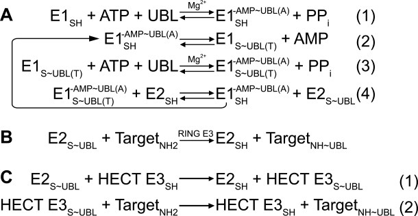
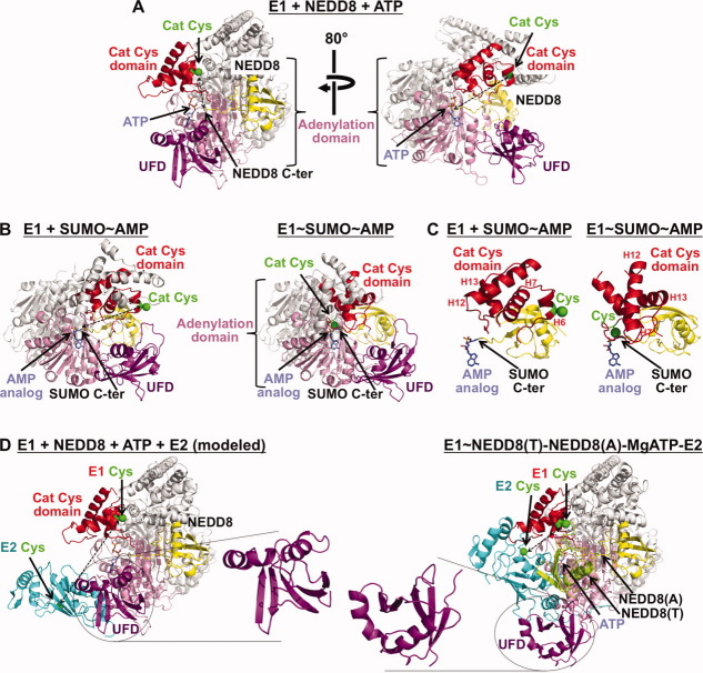
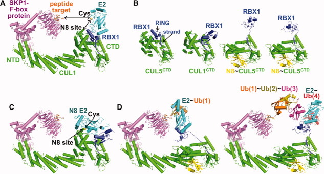
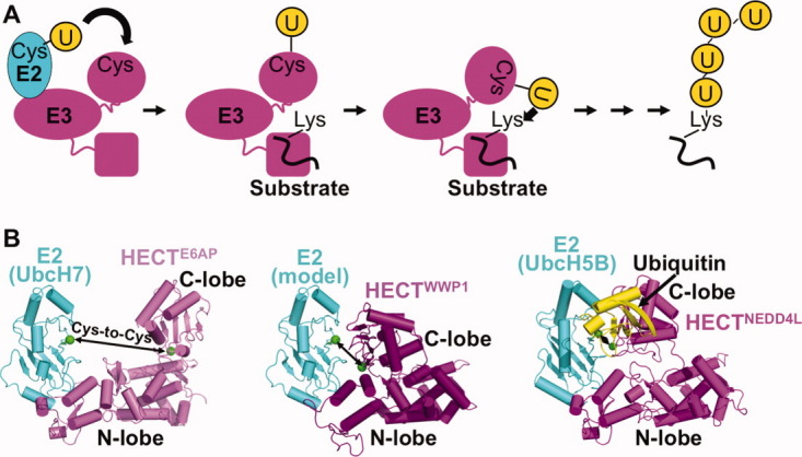
Post-translational modification by ubiquitin-like proteins (UBLs) is a predominant eukaryotic regulatory mechanism. The vast reach of this form of regulation extends to virtually all eukaryotic processes that involve proteins. UBL modifications play critical roles in controlling the cell cycle, transcription, DNA repair, stress responses, signaling, immunity, plant growth, embryogenesis, circadian rhythms, and a plethora of other pathways. UBLs dynamically modulate target protein properties including enzymatic activity, conformation, half-life, subcellular localization, and intermolecular interactions. Moreover, the enzymatic process of UBL ligation to proteins is itself dynamic, with the UBL moving between multiple enzyme active sites and ultimately to a target. This review highlights our work on how the dynamic conformations of selected enzymes catalyzing UBL ligation help mediate this fascinating form of protein regulation.


Figures

Figure 1
Figure 1
Generalized enzymatic mechanisms of UBL transfer between enzymes and ultimately to a target, based on studies of ubiquitin. ∼ refers to covalent complex; - refers to noncovalent complex. A, Initial steps catalyzed by E1. (1) E1 binds MgATP and a UBL, and catalyzes acyl-adenylation of the UBL's C-terminus. (2) E1 catalytic cysteine attacks the UBL∼AMP intermediate, to form the covalent thioester-linked E1∼UBL intermediate. (3) E1 repeats adenylation reaction on a 2nd UBL molecule, such that E1 binds 2 UBL molecules: UBL(T) is thioester-linked to E1's catalytic cystine; UBL(A) is associated noncovalently at the adenylation site. (4) Doubly-UBL-loaded E1 binds an E2. UBL(T) is transferred from the E1 cysteine to the E2 catalytic cysteine. B, RING E3s (ca. 600 in humans) enhance UBL transfer from E2 to a target. C, HECT E3s (almost 30 in humans) contain a catalytic cysteine, and (1) form a covalent thioester intermediate with a UBL prior to UBL ligation to a target lysine (2). Adapted from Ref. 38.
Figure 2
Figure 2
Dynamic assembly-line like UBL activation by E1s. Some substructures conserved in the E1s for NEDD8, SUMO, and ubiquitin are colored as follows: part of the adenylation domain is shown in pink, part of the catalytic cysteine domain is shown in red with the active site Cys in green with its side-chain as a sphere, and the E2-binding ubiquitin-fold domain (UFD) is shown in purple. Portions of the structures not discussed in the text, undergoing lesser conformational changes, or not conserved among E1s are shown in white and light grey. A, Two views, separated by an 80° rotation around the y-axis, of an E1-NEDD8-ATP complex highlighting domains that are common among E1s and that undergo significant conformational rearrangement during the activation cycle. NEDD8 is shown in yellow with its C-terminus approaching the α-phosphate of ATP. A dashed line shows the large gap between the E1 Cys and the UBL C-terminus, suggesting conformational change for forming a thioester-linked E1∼UBL intermediate. B, Chemically-trapped mimics of complexes between SUMO's E1 and SUMO after the adenylation reaction (left) and a transition-state analog for forming the covalent E1∼SUMO intermediate (right). Remarkable structural remodeling accompanies this transition, including rotation of the catalytic cysteine domain, dismantling of some substructures such as the helix that contains the catalytic cysteine during the adenylation reaction that does not involve the catalytic Cys, and formation of new catalytic substructures such as the loop containing the catalytic cysteine that is covalently linked to the UBL's C-terminus in the right structure. The structures are oriented by superimposition on the adenylation domain of NEDD8's E1 in the right panel in A. C, Same as B, but close-up views highlighting the reorientation/structural remodeling of the catalytic cysteine domain (red) of SUMO's E1 between the structures representing adenylation of SUMO (yellow) (left) and formation of the E1∼SUMO covalent intermediate (right). Helices 6, 7, 12, and 13 from SUMO's E1 are labeled H6, H7, H12, and H13, respectively, at roughly the same relative locations on the two structures, except for H6 which has “melted” into a loop in the structure mimicking the E1∼SUMO∼ AMP intermediate. D, Left, docking model of NEDD8 E2 (cyan) binding to the NEDD8 E1 UFD as in the structure in A, left panel. In docking models of E2 binding to NEDD8 or SUMO E1s, E1 and E2 cysteines (green spheres) face opposite directions and are widely separated (dashed line)., Right, structure of a trapped activation complex containing NEDD8's E1, two NEDD8s [one thioester-linked to E1—lime (T), one noncovalently associated for adenylation—yellow (A)], a catalytically inactive NEDD8 E2 (Ubc12, cyan), and MgATP. This structure revealed multiple conformational changes. First, in the double-UBL-loaded E1, the catalytic cysteine domain has returned to its original position and conformation. Second, the UFD (purple, highlighted) has undergone significant rotation such that the E2 cysteine now faces the E1.
Figure 3
Figure 3
Examples of RING domain rotation and conformational control of cullin-RING E3 ligases. A, Initial structural model of an SCF CRL (SKP1 and the F-box protein β-TRCP in purple, CUL1 in green, RBX1 in blue) bound to the peptide target (β-catenin phosphopeptide, orange sticks) and a generic E2 (cyan, with catalytic cysteine in yellow). Gaps between E2 cysteine and CUL1's NEDD8 acceptor lysine (“N8 site,” spheres) and to the ubiquitination target (in this case, peptide) are indicated with arrows.,,, B, RBX1 RING domain orientational flexibility revealed from comparing structures of CUL5CTD-RBX1 (RBX1 RING in a similar orientation as in CUL1-RBX1 as in panel A), CUL1CTD-RBX1 with the RBX1 RING domain in a different orientation, and two complexes of NEDD8∼ CUL5CTD-RBX1, with cullin C-terminal domains (CTDs) in green, RBX1 in blue, and NEDD8 in yellow. Structures are aligned over the subdomain containing the RBX1 strand. C, Model for juxtaposition of the CUL1 NEDD8 acceptor lysine (N8 site) and NEDD8 E2 catalytic cysteine based on docking NEDD8's E2 Ubc12 (cyan with catalytic Cys in yellow) onto SCFβTRCP as in A but substituted with RBX1 from the CUL1CTD-RBX1 as in B, with the RBX1 RING domain in an orientation poised for NEDD8 ligation.,,,,, D, Model for ubiquitin (orange) transfer from ubiquitin E2 (cyan) to the ubiquitination peptide target by a NEDD8-activated CRL. The E2 cysteine and peptide target are first brought into proximity via extension/rotation of the RBX1 linker (left), with polyubiquitination (ubiquitins in orange, olive, magenta and red) by a NEDD8-activated CRL, where rotation about the RBX1 linker allows multiple catalytic geometries associated with building a polyubiquitin chain.
Figure 4
Figure 4
HECT E3 conformational dynamics. A, Schematic view of HECT E3 ubiquitin transfer mechanism. HECT E3s have an N-terminal region of varying sequence/structure containing protein interaction domains responsible for substrate binding and subcellular localization, and a C-terminal HECT domain. The HECT domain contains two “lobes,” an “N-lobe” that binds an E2 and a “C-lobe” that contains the catalytic cysteine. Ubiquitin is first transferred from an associated E2 to the HECT catalytic cysteine, and then to a lysine residue on the target. It is thought that conformational changes accompany many steps in HECT E3-mediated ubiquitin transfer. B, Structural insights into the first HECT E3 function of E2-to-E3 ubiquitin transfer. Shown from left to right are E6AP HECT domain (violet) bound to the E2 UbcH7 (cyan), WWP1 HECT domain (purple) with UbcH7 (cyan) modeled, and NEDD4L HECT domain (magenta) bound to UbcH5B (cyan)∼ubiquitin (yellow). Arrows denote gaps between the E2 and HECT domain cysteines (green). The structures were aligned by superimposing the HECT domain N-lobes.

References

    1. Hicke L. Protein regulation by monoubiquitin. Nat Rev. 2001;2:195–201. - PubMed
    1. Hurley JH, Stenmark H. Molecular mechanisms of ubiquitin-dependent membrane traffic. Annu Rev Biophys. 2011;40:119–142. - PMC - PubMed
    1. Di Fiore PP, Polo S, Hofmann K. When ubiquitin meets ubiquitin receptors: a signalling connection. Nature reviews. Mol Cell Biol. 2003;4:491–497. - PubMed
    1. Hicke L, Schubert HL, Hill CP. Ubiquitin-binding domains. Nat Rev. 2005;6:610–621. - PubMed
    1. Morita E, Sundquist WI. Retrovirus budding. Ann Rev Cell Dev Biol. 2004;20:395–425. - PubMed

Publication types

MeSH terms

LinkOut - more resources