

Percutaneous ablation in the kidney
- PMID: 22012904
- PMCID: PMC3198222
- DOI: 10.1148/radiol.11091207
Percutaneous ablation in the kidney
Abstract
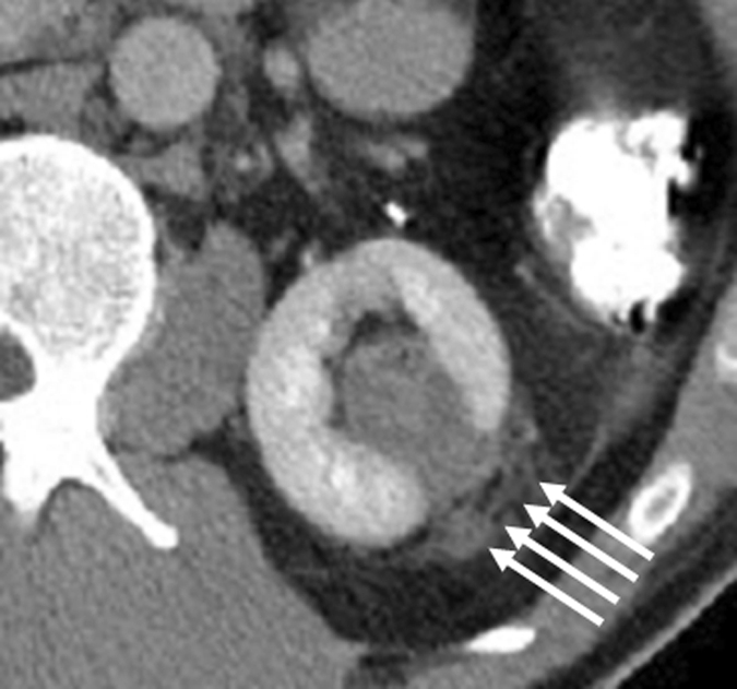
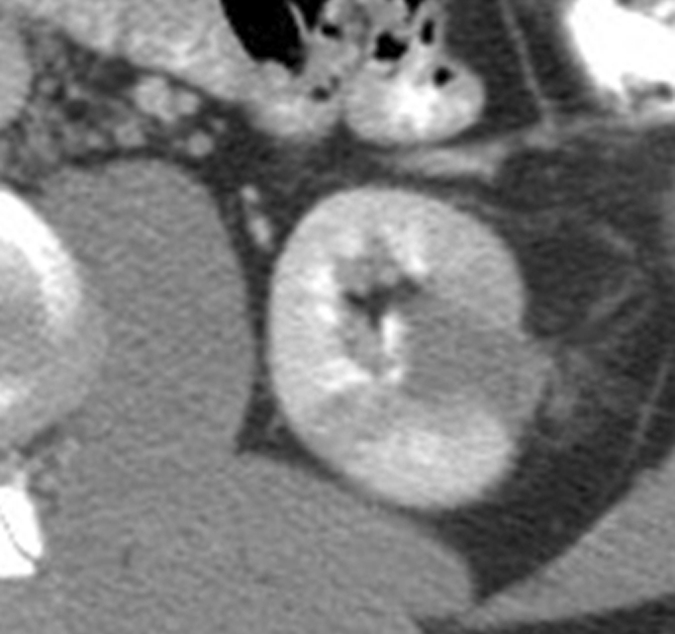
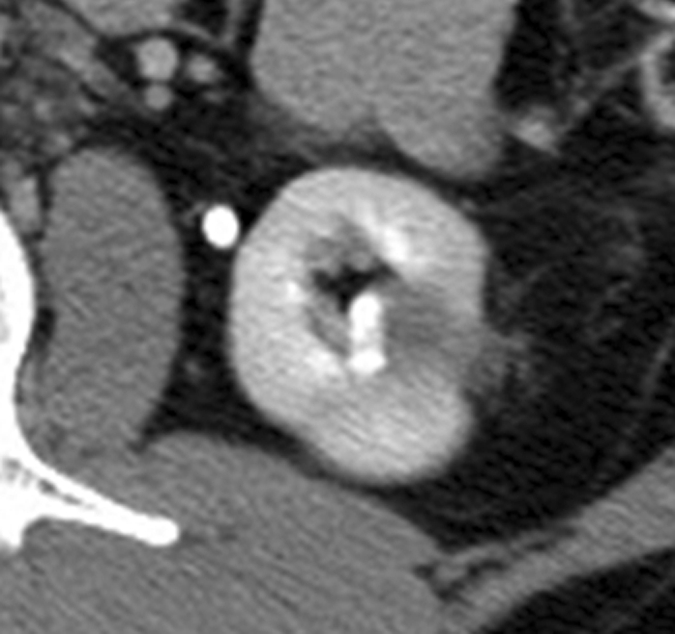
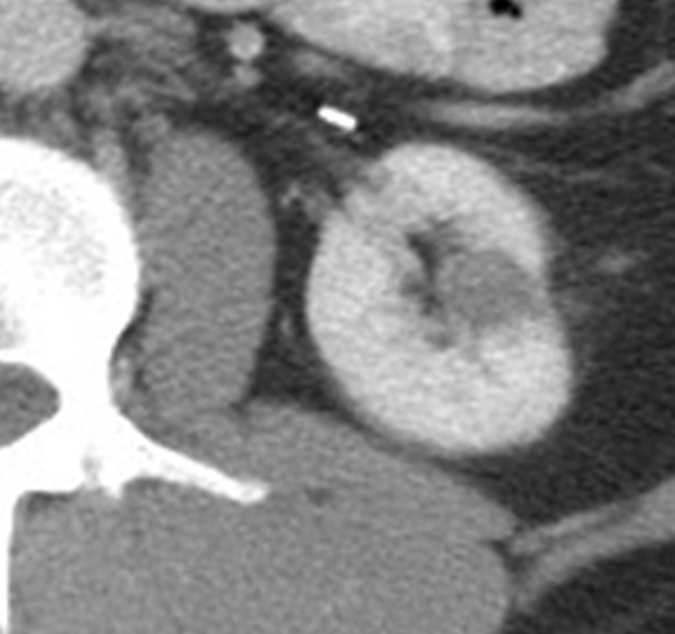
Percutaneous ablation in the kidney is now performed as a standard therapeutic nephron-sparing option in patients who are poor candidates for resection. Its increasing use has been largely prompted by the rising incidental detection of renal cell carcinomas with cross-sectional imaging and the need to preserve renal function in patients with comorbid conditions, multiple renal cell carcinomas, and/or heritable renal cancer syndromes. Clinical studies to date indicate that radiofrequency ablation and cryoablation are effective therapies with acceptable short- to intermediate-term outcomes and with a low risk in the appropriate setting, with attention to pre-, peri-, and postprocedural detail. The results following percutaneous radiofrequency ablation and cryoablation in the treatment of renal cell carcinoma are reviewed in this article, including those of several larger scale studies of ablation of T1a tumors. Clinical and technical considerations unique to ablation in the kidney are presented, and potential complications are discussed.
RSNA, 2011
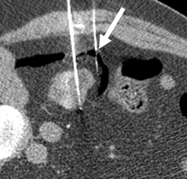
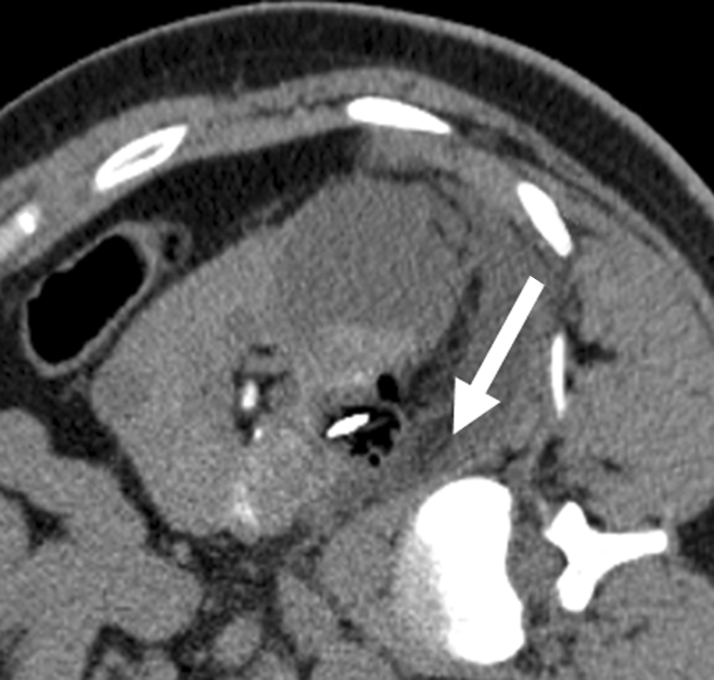
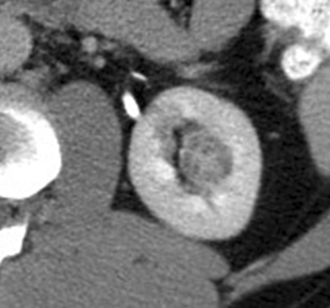
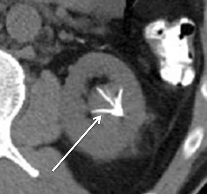
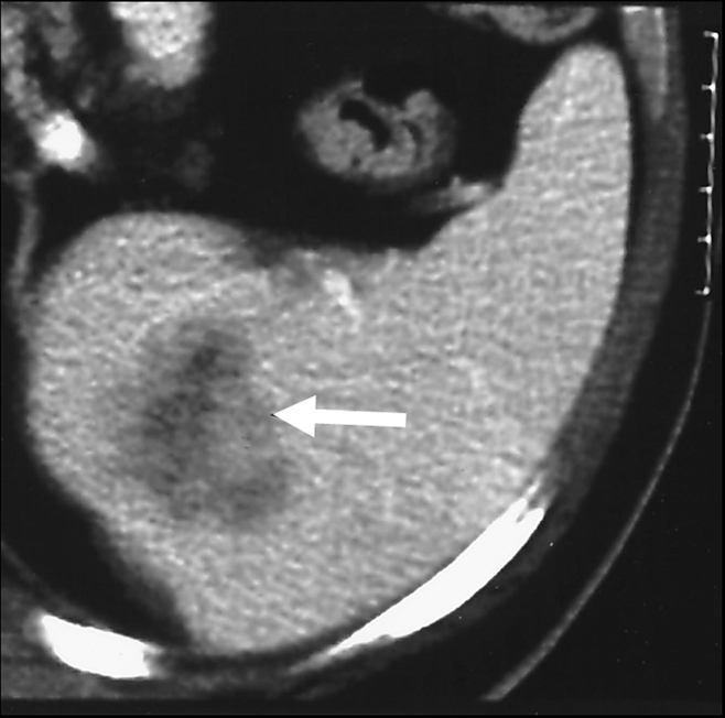
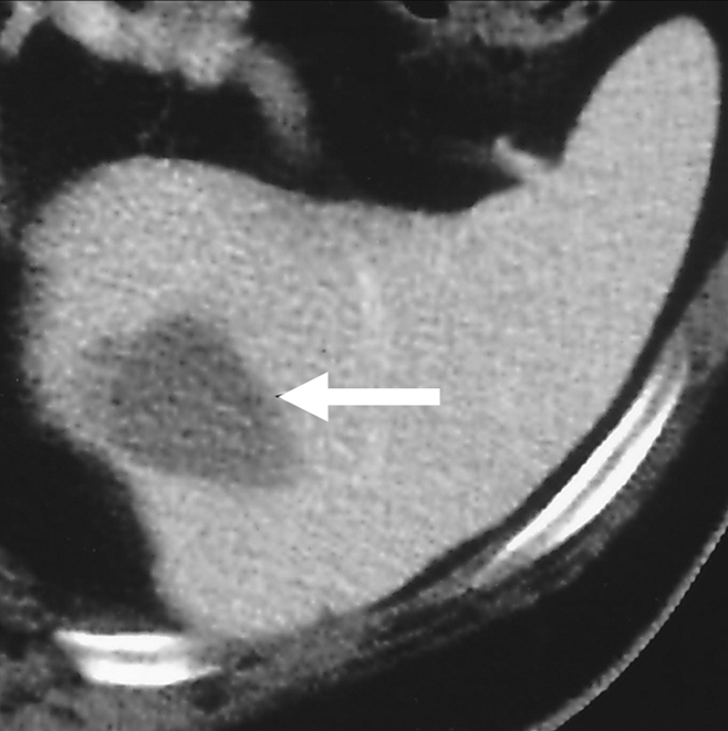
Figures
Comment in
-
Re: Percutaneous ablation in the kidney.J Urol. 2012 Apr;187(4):1224-5. doi: 10.1016/j.juro.2011.12.048. Epub 2012 Feb 14. J Urol. 2012. PMID: 22423905 No abstract available.
References
-
- Cohen HT, McGovern FJ. Renal-cell carcinoma. N Engl J Med 2005;353(23):2477–2490 - PubMed
-
- National Cancer Institute Kidney cancer home page. http://www.cancer.gov/cancertopics/types/kidney. Accessed September 4, 2011
-
- Pantuck AJ, Zisman A, Belldegrun AS. The changing natural history of renal cell carcinoma. J Urol 2001;166(5):1611–1623 - PubMed
-
- Gervais DA, McGovern FJ, Arellano RS, McDougal WS, Mueller PR. Radiofrequency ablation of renal cell carcinoma. I. Indications, results, and role in patient management over a 6-year period and ablation of 100 tumors. AJR Am J Roentgenol 2005;185(1):64–71 - PubMed
-
- Zagoria RJ, Hawkins AD, Clark PE, et al. Percutaneous CT-guided radiofrequency ablation of renal neoplasms: factors influencing success. AJR Am J Roentgenol 2004;183(1):201–207 - PubMed
Publication types
MeSH terms
Substances
LinkOut - more resources
Full Text Sources
Medical
