. 2012 Jan;86(1):203-13.
doi: 10.1128/JVI.05817-11. Epub 2011 Oct 19.

The vaccinia virus-encoded Bcl-2 homologues do not act as direct Bax inhibitors

Affiliations

The vaccinia virus-encoded Bcl-2 homologues do not act as direct Bax inhibitors

Antonio Postigo et al. J Virol. 2012 Jan.

Abstract

Many viruses, including members of several poxvirus genera, encode inhibitors that block apoptosis by simultaneously binding the proapoptotic Bcl-2 proteins Bak and Bax. The Orthopoxvirus vaccinia virus encodes the Bcl-2-like F1 protein, which sequesters Bak but not Bax. However, N1, a potent virulence factor, is reported to be antiapoptotic and to interact with Bax. Here we investigated whether vaccinia virus inhibits Bak/Bax-dependent apoptosis via the cooperative action of F1 and N1. We found that Western Reserve (WR) and ΔN1L viruses inhibited drug- and infection-induced apoptosis equally. Meanwhile, infections with ΔF1L or ΔN1L/F1L virus resulted in similar levels of Bax activation and apoptosis. Outside the context of infection, N1 did not block drug- or Bax-induced cell death or interact with Bax. In addition to F1 and N1, vaccinia virus encodes further structural homologs of Bcl-2 proteins that are conserved in orthopoxviruses, including A46, A52, B14, C1, C6, C16/B22, K7, and N2. However, we found that these do not associate with Bax or inhibit drug-induced cell death. Based on our findings that N1 is not an antiapoptotic protein, we propose that the F1 orthologs represent the only orthopoxvirus Bcl-2 homolog to directly inhibit the Bak/Bax checkpoint.


Figures
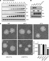
Fig 1
Fig 1
Loss of N1 does not decrease plaque size in the absence of F1. (A) Western blot analysis of whole-cell lysates of WR-infected cells probed with antibodies against F1, N1, A36 (early viral protein), A27 (late viral protein), and actin at the indicated times postinfection. (B) Western blot analysis of HeLa cell lysates infected with the indicated viruses for 20 h reveals the absence of N1 and F1 proteins after deletion of the indicated genes. (C) Representative images of plaque formation by the indicated viruses in BSC-1 cells visualized with crystal violet at 4 days postinfection. Quantitative analysis of plaque sizes of the indicated viruses shows a reduced plaque size in ΔVGF/F1L virus-infected cells. The error bars represent standard errors of the means (n = 125 for each virus). ***, P < 0.001.
Fig 2
Fig 2
F1, but not N1, inhibits apoptosis. (A) HeLa cells were infected for 5 h with the indicated viruses at an MOI of 2 before treatment with 1 μM STS for 2 and 4 h. Western blot analysis of whole-cell lysates by using antibodies against PARP, F1, N1, and tubulin is shown. (B) Western blot analysis of whole-cell lysates from floating and adherent HeLa cells infected with the indicated viruses for 20 h at an MOI of 2, using antibodies against PARP, caspase-3, A27, and tubulin. (C) Quantitative analysis of nuclear condensation in HeLa cells infected with the indicated viruses for 20 h. At least 250 cells were counted from two independent coverslips. Averages were determined for three independent experiments, and error bars represent standard errors of the means. ***, P < 0.001. (D) Quantitative immunofluorescence analysis of the percentage of HeLa cells exhibiting cytosolic cytochrome c after infection for 20 h with the indicated viruses. At least 250 cells were counted in 3 independent experiments, and error bars represent standard errors of the means. ***, P < 0.001. (E) Representative immunofluorescence images of HeLa cells infected with the indicated viruses and stained with cytochrome c antibody. Arrows illustrate cells with cytoplasmic cytochrome c.
Fig 3
Fig 3
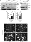
Loss of N1 does not increase Bax activation during infection. (A) Representative immunofluorescence images of HeLa cells infected for 20 h with the indicated viruses and stained with antibody (6A7) to active Bax. The appearance of activated Bax was observed predominantly in cells infected with the ΔF1L and ΔN1L/F1L viruses. (B) Quantitative analysis of Bax activation in cells infected for 20 h with the indicated viruses. The percentage of cells exhibiting staining of active Bax was determined for at least 250 cells in three independent experiments. Error bars represent standard errors of the means. ***, P < 0.001. (C) Western blot analysis reveals that activated Bax can be immunoprecipitated (IP) readily from HeLa cells infected with the ΔF1L and ΔN1L/F1L viruses but not the WR and ΔN1L viruses.
Fig 4
Fig 4
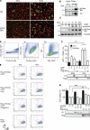
N1 expression does not inhibit Bax-induced cell death. (A) Representative immunofluorescence images of HeLa-T-Rex-HA-Bax cells stained with antibodies to HA (top) or active Bax (bottom) (green), before (left) or after (right) induction with doxycycline. Cells were highlighted by nuclear staining with Hoechst 33342 (red). (B) Western blot analysis of HeLa-T-Rex-empty vector (E.V.) and -HA-Bax cell lysates showing that HA-Bax is induced by doxycycline addition for 20 h. (C) Western blot analysis of doxycycline-induced HA-Bax expression and PARP cleavage in HeLa-T-Rex-HA-Bax cells over 24 h. (D) Gating strategy used in flow cytometry analysis of apoptosis. Panel I shows a region that includes all singlet events, based on their side scatter width value. This region was then applied to panel II, and whole cells were gated as shown, on the basis of their forward and side scatter signals. This cell population was then used in panel III to define GFP-positive cells. (E) Each plot displays only events that satisfy all three criteria in panel D. For each cell line and condition, TMRE fluorescence (x axis) is shown against DAPI fluorescence (y axis). Live cells are defined as TMREhigh and DAPI negative, apoptotic cells are TMRElow and DAPI negative, and dead cells are DAPI positive. Regions were set using the HeLa-GFP control and applied to all other plots. (F) Quantitative flow cytometry analysis of apoptosis in HeLa-T-Rex-empty vector (black bars) and HeLa-T-Rex-HA-Bax (white bars) cells expressing the indicated proteins for 24 h followed by induction with doxycycline for 20 h. Apoptosis was assessed by mitochondrial dysfunction analysis using TMRE fluorescence and was quantified as the percentage of GFP-positive cells that were both TMRE and DAPI negative. Western blots show the expression of N1 and Flag-M11L under the different conditions. Error bars represent standard errors of the means for 3 independent experiments. **, P < 0.01. (G) Cell viability analysis of HeLa cells transfected with increasing amounts of HA-Bax plasmid and constant amounts of empty vector or N1L or Flag-M11L plasmid (in nanograms), as indicated. Western blots show the presence of N1 and Flag-M11L in the lysates derived from the remaining adherent cells. The data represent means ± standard errors of the means for three independent experiments performed in quadruplicate. **, P < 0.01; ***, P < 0.001.
Fig 5
Fig 5
N1 does not interact with Bax. (A and B) HeLa-T-Rex-HA-Bax cells were induced with doxycycline for 20 h and then infected with WR for 6 h. Immunoprecipitation of N1 with mouse monoclonal anti-N1 (A) and anti-active Bax agarose (0.N.18) (B) was performed with infected cell lysates. Western blots of the immunoprecipitates and the whole-cell lysates (input) were probed using anti-Bax (Bax NT) and polyclonal anti-N1 antibodies. (C and D) HEK 293 cells infected with WR for 16 h at an MOI of 2 were lysed and subjected to immunoprecipitation with mouse monoclonal GFP or N1 antibody. Western blots of the immunoprecipitates and the cell lysates (input) were probed using antibodies against Bad (C) and Bid (D).
Fig 6
Fig 6
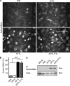
N1 does not inhibit STS-induced cell death. (A) Western blot analysis of cell lysates from HEK293-T-Rex-empty vector (E.V.) and -N1L cells showing the level of N1 induction after 20 h of doxycycline treatment. (B) Representative immunofluorescence images of HEK293-T-Rex-E.V. (left) and HEK293-T-Rex-N1L (right) cells stained with monoclonal anti-N1 antibody (green), before (top) or after (bottom) induction with doxycycline for 20 h. Cells were highlighted by nuclear staining with Hoechst 33342 (red). (C) Western blot analysis of cell lysates from HEK293-T-Rex-empty vector (E.V.) and -N1L cells treated with doxycycline or vehicle for 20 h, followed by UV irradiation at 200 mJ/cm2. Cell lysates were prepared 4 h after irradiation, and apoptosis induction was assessed by Western blot analysis of PARP cleavage. (D) Cell viability analysis of HEK293-T-Rex-E.V. (left) and HEK293-T-Rex-N1L (right) cells induced or not with doxycycline for 20 h and treated for 6 h with increasing concentrations of STS. Black bars show cells not treated with STS. Graphs show data from a representative experiment, with each bar showing the mean and standard error of the mean for quintuplicate samples.
Fig 7
Fig 7
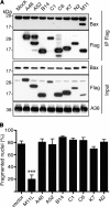
Vaccinia virus Bcl-2 homologs do not interact with Bax or inhibit apoptosis. (A) HeLa cells infected with WR were transfected with the indicated pE/L-Flag constructs at 2 hpi and lysed 18 h later. Western blots of immunoprecipitates obtained using Flag antibody agarose (M2) and whole-cell lysates (input) were probed with antibodies against Bax, N1, and A36. The asterisk denotes the antibody light chain. (B) HeLa cells transfected with pEGFP and the indicated pCDNA4-Flag constructs were treated at 24 h posttransfection with STS for 4 h. The percentage of apoptosis was determined by quantification of fragmented nuclei in at least 150 GFP-positive cells in three independent experiments. Error bars represent standard errors of the means. ***, P < 0.001.

Similar articles

Cited by

References

    1. Adams JM, Cory S. 2007. Bcl-2-regulated apoptosis: mechanism and therapeutic potential. Curr. Opin. Immunol. 19:488–496 - PMC - PubMed
    1. Afonso CL, et al. 2000. The genome of fowlpox virus. J. Virol. 74:3815–3831 - PMC - PubMed
    1. Aoyagi M, et al. 2007. Vaccinia virus N1L protein resembles a B cell lymphoma-2 (Bcl-2) family protein. Protein Sci. 16:118–124 - PMC - PubMed
    1. Arakawa Y, Cordeiro JV, Way M. 2007. F11L-mediated inhibition of RhoA-mDia signaling stimulates microtubule dynamics during vaccinia virus infection. Cell Host Microbe 1:213–226 - PubMed
    1. Arnoult D, et al. 2004. Cytomegalovirus cell death suppressor vMIA blocks Bax- but not Bak-mediated apoptosis by binding and sequestering Bax at mitochondria. Proc. Natl. Acad. Sci. U. S. A. 101:7988–7993 - PMC - PubMed

Publication types

MeSH terms

Substances