

doi: 10.1128/JVI.05151-11.
Epub 2011 Oct 19.
Epstein-Barr virus isolates retain their capacity to evade T cell immunity through BNLF2a despite extensive sequence variation
Affiliations
- PMID: 22013037
- PMCID: PMC3255881
- DOI: 10.1128/JVI.05151-11
Item in Clipboard
Epstein-Barr virus isolates retain their capacity to evade T cell immunity through BNLF2a despite extensive sequence variation
J Virol.
2012 Jan.
Abstract
The Epstein-Barr virus (EBV)-encoded immune evasion protein BNLF2a inhibits the transporter associated with antigen processing (TAP), thereby downregulating HLA class I expression at the cell surface. As a consequence, recognition of EBV-infected cells by cytotoxic T cells is impaired. Here, we show that sequence polymorphism of the BNLF2a protein is observed with natural EBV isolates, with evidence for positive selection. Despite these mutations, the BNLF2a variants efficiently reduce cell surface HLA class I levels. This conservation of BNLF2a function during evolution of EBV implies an important role for the viral TAP inhibitor in preventing T cell recognition during viral infection.
Figures
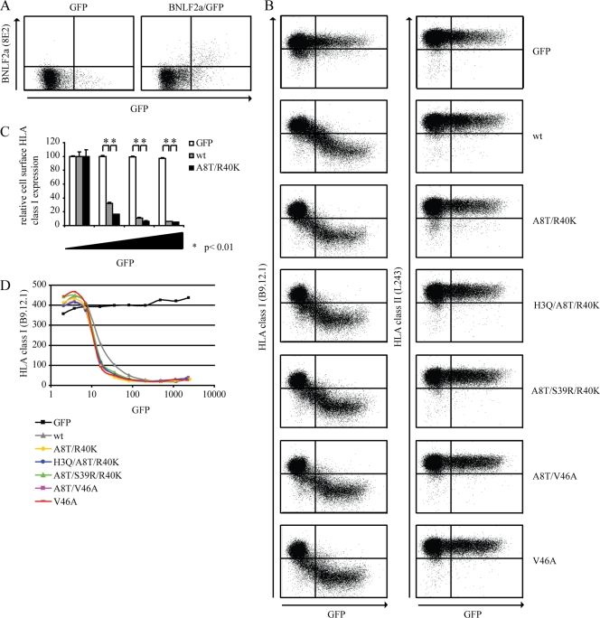
EBV isolates retain BNLF2a-mediated HLA I downregulation. (A) MJS cells were transiently transfected to express the control protein GFP or to coexpress wild-type BNLF2a and GFP. After 48 h, cells were stained for intracellular expression of BNLF2a (monoclonal antibody [MAb] 8E2). Subsequently, the cells were analyzed by flow cytometry using CellQuest Pro software (BD Biosciences). (B) MJS cells were transiently transfected to express the control protein GFP, wild-type BNLF2a (wt), or one of the following BNLF2a mutants: A8T/R40K, H3Q/A8T/R40K, A8T/S39R/R40K, A8T/V46A, or V46A. After 48 h, cells were stained for cell surface expression of HLA I (MAb B9.12.1) and HLA II (MAb L243) and analyzed by flow cytometry using CellQuest Pro software (BD Biosciences). (C) Quantification of flow cytometry data. Cell surface expression levels of HLA I were correlated with GFP expression for cells transfected to express the control protein GFP, wild-type (wt) BNLF2a, or the A8T/R40K BNLF2a mutant. To this end, values were corrected for cell surface expression of HLA I in GFP-negative cells. The standard deviations are represented by the error bars. *, P < 0.01 as determined by a t test. (D) Graphical display of the results shown in panel B. The mean fluorescence index of HLA I expression is plotted against the mean fluorescence index of GFP expression. The results of one representative experiment out of at least three independent experiments are shown. For panel A, the experiment was performed in duplicate.
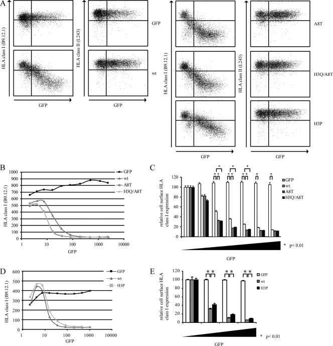
N-terminal amino acids of BNLF2a affect its immune evasion function. (A) MJS cells were transiently transfected to express the control protein GFP, wild-type BNLF2a (wt), BNLF2a-A8T (A8T), BNLF2a-H3Q/A8T (H3Q/A8T), or BNLF2a-H3P (H3P). After 48 h, cells were stained for cell surface expression of HLA I (MAb B9.12.1) and HLA II (MAb L243) and analyzed by flow cytometry using CellQuest Pro software (BD Biosciences). (B and D) Graphical display of flow cytometry data. The mean fluorescence index of HLA I expression is plotted against the mean fluorescence index of GFP expression. (C and E) Quantification of flow cytometry data. Cell surface expression levels of HLA I were correlated with GFP expression. To this end, values were corrected for cell surface expression of HLA I in GFP-negative cells. The standard deviations are represented by the error bars. *, P < 0.01 as determined by a t test. The results of one representative experiment out of three independent experiments are shown.
Similar articles
-
Stage-specific inhibition of MHC class I presentation by the Epstein-Barr virus BNLF2a protein during virus lytic cycle.PLoS Pathog. 2009 Jun;5(6):e1000490. doi: 10.1371/journal.ppat.1000490. Epub 2009 Jun 26. PLoS Pathog. 2009. PMID: 19557156 Free PMC article.
-
Latent Expression of the Epstein-Barr Virus (EBV)-Encoded Major Histocompatibility Complex Class I TAP Inhibitor, BNLF2a, in EBV-Positive Gastric Carcinomas.J Virol. 2015 Oct;89(19):10110-4. doi: 10.1128/JVI.01110-15. Epub 2015 Jul 15. J Virol. 2015. PMID: 26178981 Free PMC article.
-
EBV protein BNLF2a exploits host tail-anchored protein integration machinery to inhibit TAP.J Immunol. 2011 Mar 15;186(6):3594-605. doi: 10.4049/jimmunol.1002656. Epub 2011 Feb 4. J Immunol. 2011. PMID: 21296983
-
Epstein-Barr virus evasion of CD8(+) and CD4(+) T cell immunity via concerted actions of multiple gene products.Semin Cancer Biol. 2008 Dec;18(6):397-408. doi: 10.1016/j.semcancer.2008.10.008. Epub 2008 Oct 25. Semin Cancer Biol. 2008. PMID: 18977445 Review.
-
Contribution of viral recombinants to the study of the immune response against the Epstein-Barr virus.Semin Cancer Biol. 2008 Dec;18(6):409-15. doi: 10.1016/j.semcancer.2008.09.001. Epub 2008 Sep 30. Semin Cancer Biol. 2008. PMID: 18938248 Review.
Cited by
-
Immune control of oncogenic γ-herpesviruses.Curr Opin Virol. 2015 Oct;14:79-86. doi: 10.1016/j.coviro.2015.08.014. Epub 2015 Sep 13. Curr Opin Virol. 2015. PMID: 26372881 Free PMC article. Review.
-
Categorizing Sequences of Concern by Function To Better Assess Mechanisms of Microbial Pathogenesis.Infect Immun. 2022 May 19;90(5):e0033421. doi: 10.1128/IAI.00334-21. Epub 2021 Nov 15. Infect Immun. 2022. PMID: 34780277 Free PMC article. Review.
-
Novel Therapies Boosting T Cell Immunity in Epstein Barr Virus-Associated Nasopharyngeal Carcinoma.Int J Mol Sci. 2020 Jun 16;21(12):4292. doi: 10.3390/ijms21124292. Int J Mol Sci. 2020. PMID: 32560253 Free PMC article. Review.
-
An Epstein-Barr virus protein interaction map reveals NLRP3 inflammasome evasion via MAVS UFMylation.Mol Cell. 2023 Jul 6;83(13):2367-2386.e15. doi: 10.1016/j.molcel.2023.05.018. Epub 2023 Jun 12. Mol Cell. 2023. PMID: 37311461 Free PMC article.
-
An Epstein-Barr Virus MicroRNA Blocks Interleukin-1 (IL-1) Signaling by Targeting IL-1 Receptor 1.J Virol. 2017 Oct 13;91(21):e00530-17. doi: 10.1128/JVI.00530-17. Print 2017 Nov 1. J Virol. 2017. PMID: 28794034 Free PMC article.
References
-
- Apolloni A, et al. 1992. Sequence variation of cytotoxic T cell epitopes in different isolates of Epstein-Barr virus. Eur. J. Immunol. 22:183–189 - PubMed
-
- Baer R, et al. 1984. DNA sequence and expression of the B95-8 Epstein-Barr virus genome. Nature 310:207–211 - PubMed
-
- Bell MJ, et al. 2008. Widespread sequence variation in Epstein-Barr virus nuclear antigen 1 influences the antiviral T cell response. J. Infect. Dis. 197:1594–1597 - PubMed
Publication types
MeSH terms
Substances
Associated data
- Actions
- Actions
- Actions
- Actions
- Actions
- Actions
- Actions
- Actions
- Actions
- Actions
- Actions
- Actions
- Actions
- Actions
- Actions
- Actions
- Actions
- Actions
- Actions
- Actions
- Actions
- Actions
- Actions
- Actions
- Actions
- Actions
- Actions
- Actions
- Actions
- Actions
- Actions
- Actions
- Actions
- Actions
- Actions
- Actions
- Actions
- Actions
- Actions
- Actions
- Actions
- Actions
- Actions
- Actions
- Actions
- Actions
- Actions
- Actions
- Actions
- Actions
- Actions
- Actions
- Actions
Grants and funding
LinkOut - more resources
Full Text Sources
Research Materials
Miscellaneous
