. 2012 Jan;86(1):114-20.
doi: 10.1128/JVI.05590-11. Epub 2011 Oct 19.

Latent HIV-1 infection of resting CD4⁺ T cells in the humanized Rag2⁻/⁻ γc⁻/⁻ mouse

Affiliations

Latent HIV-1 infection of resting CD4⁺ T cells in the humanized Rag2⁻/⁻ γc⁻/⁻ mouse

Shailesh K Choudhary et al. J Virol. 2012 Jan.

Abstract

Persistent human immunodeficiency virus type 1 (HIV-1) infection of resting CD4⁺ T cells, unaffected by antiretroviral therapy (ART), provides a long-lived reservoir of HIV infection. Therapies that target this viral reservoir are needed to eradicate HIV-1 infection. A small-animal model that recapitulates HIV-1 latency in resting CD4⁺ T cells may accelerate drug discovery and allow the rational design of nonhuman primate (NHP) or human studies. We report that in humanized Rag2⁻/⁻ γ(c)⁻/⁻ (hu-Rag2⁻/⁻ γ(c)⁻/⁻) mice, as in humans, resting CD4⁺ T cell infection (RCI) can be quantitated in pooled samples of circulating cells and tissue reservoirs (e.g., lymph node, spleen, bone marrow) following HIV-1 infection with the CCR5-tropic JR-CSF strain and suppression of viremia by ART. Replication-competent virus was recovered from pooled resting CD4⁺ T cells in 7 of 16 mice, with a median frequency of 8 (range, 2 to 12) infected cells per million T cells, demonstrating that HIV-1 infection can persist despite ART in the resting CD4⁺ T cell reservoir of hu-Rag2⁻/⁻ γ(c)⁻/⁻ mice. This model will allow rapid preliminary assessments of novel eradication approaches and combinatorial strategies that may be challenging to perform in the NHP model or in humans, as well as a rigorous analysis of the effect of these interventions in specific anatomical compartments.


Figures

Fig 1
Fig 1
Characterization of human CD4+ (huCD4+) T cell reconstitution in lymphoid tissues and peripheral blood (PB). Cells from mesenteric lymph nodes, bone marrow, and spleen were harvested at 14 weeks posttransplantation and characterized by flow cytometric analysis. Live cells were discriminated based on forward and side scatter and gated on human CD45+/mouse CD45 cells to quantitate human-cell reconstitution in each tissue (shown in parentheses). (A) Human CD45+ cells were gated on CD3+ cells to analyze CD4/CD8 T cells. (B and C) CD4+ T cells were characterized as naïve or memory cells based on expression of CD62L or the CD45RO antigen, respectively. (C and D) The resting state of CD4+ cells was determined by the lack of activation markers CD25, CD69, and HLA-DR. Data are representative of results from 5 uninfected animals.
Fig 2
Fig 2
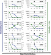
Suppression of HIV-1 plasma viremia with ART in hu-Rag2−/− γc−/− mice. Mice were infected with JR-CSF (25 ng p24/mouse) and treated with the nucleoside/nucleotide reverse transcriptase inhibitors emtricitabine (FTC) and tenofovir (TFV), the integrase strand transfer inhibitor L-870812, and the fusion inhibitor enfuvirtide (60, 60, 20, and 100 mg/kg/daily, respectively). BI, time before infection. The arrows indicate the initiation of ART, the blue lines and triangles show plasma viremia, while the green lines and squares show the percentages of all PBMCs that are human CD3+ CD4+ T cells. The blue dotted lines show the limit of detection (<800 copies/ml) of plasma viremia in 50 to 100 μl of samples obtained through the tail vein. The filled markers below the dotted lines indicate detection of plasma viremia in higher sample volumes at necropsy (limit of detection, 40 copies/ml).
Fig 3
Fig 3
Isolation of resting CD4+ T cells from humanized mice. Resting CD4+ T cells were isolated from pooled blood and lymphoid tissues of each mouse by negative selection. Cells before (A) and after (B) magnetic bead enrichment are shown. CD4+ T cells lacked the activation markers CD25 and HLA-DR and were predominantly central memory cells (CD27+ CD45RA+). mCD45, mouse CD45+ cells.

References

    1. Anderson JA, et al. 2011. Clonal sequences recovered from plasma from patients with residual HIV-1 viremia and on intensified antiretroviral therapy are identical to replicating viral RNAs recovered from circulating resting CD4+ T cells. J. Virol. 85:5220–5223 - PMC - PubMed
    1. Archin NM, et al. 2008. Valproic acid without intensified antiviral therapy has limited impact on persistent HIV infection of resting CD4+ T cells. AIDS 22:1131–1135 - PMC - PubMed
    1. Archin NM, et al. 2009. Expression of latent HIV induced by the potent HDAC inhibitor suberoylanilide hydroxamic acid. AIDS Res. Hum. Retroviruses 25:207–212 - PMC - PubMed
    1. Baenziger S, et al. 2006. Disseminated and sustained HIV infection in CD34+ cord blood cell-transplanted Rag2−/−gamma c−/− mice. Proc. Natl. Acad. Sci. U. S. A. 103:15951–15956 - PMC - PubMed
    1. Bailey JR, et al. 2006. Residual human immunodeficiency virus type 1 viremia in some patients on antiretroviral therapy is dominated by a small number of invariant clones rarely found in circulating CD4+ T cells. J. Virol. 80:6441–6457 - PMC - PubMed

Publication types

MeSH terms