. 2012 Jan;86(1):406-19.
doi: 10.1128/JVI.05720-11. Epub 2011 Oct 19.

Secretion modification region-derived peptide disrupts HIV-1 Nef's interaction with mortalin and blocks virus and Nef exosome release

Affiliations

Secretion modification region-derived peptide disrupts HIV-1 Nef's interaction with mortalin and blocks virus and Nef exosome release

Martin N Shelton et al. J Virol. 2012 Jan.

Abstract

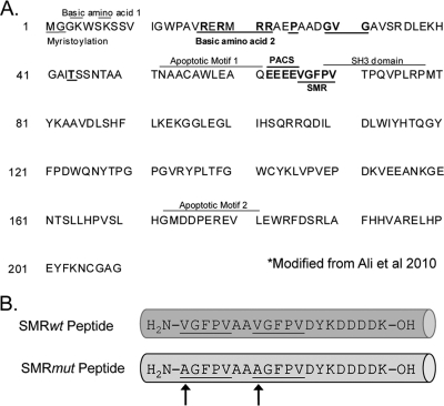
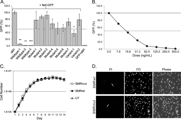
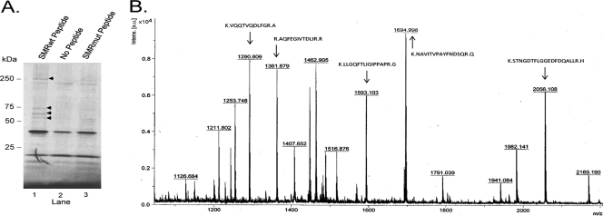
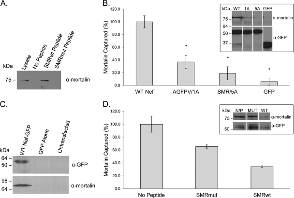
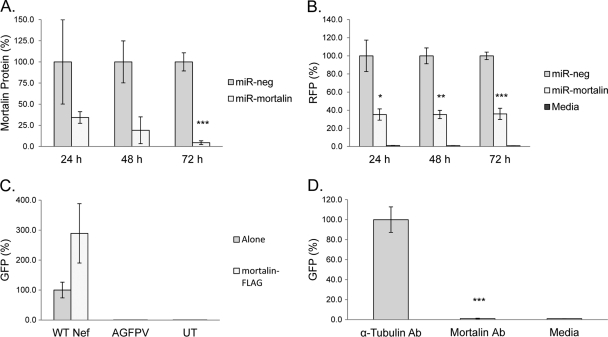
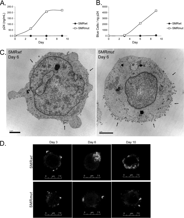
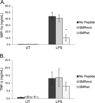
Nef is secreted from infected cells in exosomes and is found in abundance in the sera of HIV-infected individuals. Secreted exosomal Nef (exNef) induces apoptosis in uninfected CD4⁺ T cells and may be a key component of HIV pathogenesis. The exosomal pathway has been implicated in HIV-1 virus release, suggesting a possible link between these two viral processes. However, the underlying mechanisms and cellular components of exNef secretion have not been elucidated. We have previously described a Nef motif, the secretion modification region (SMR; amino acids 66 to 70), that is required for exNef secretion. In silico modeling data suggest that this motif can form a putative binding pocket. We hypothesized that the Nef SMR binds a cellular protein involved in protein trafficking and that inhibition of this interaction would abrogate exNef secretion. By using tandem mass spectrometry and coimmunoprecipitation with a novel SMR-based peptide (SMRwt) that blocks exNef secretion and HIV-1 virus release, we identified mortalin as an SMR-specific cellular protein. A second set of coimmunoprecipitation experiments with full-length Nef confirmed that mortalin interacts with Nef via Nef's SMR motif and that this interaction is disrupted by the SMRwt peptide. Overexpression and microRNA knockdown of mortalin revealed a positive correlation between exNef secretion levels and mortalin protein expression. Using antibody inhibition we demonstrated that the Nef/mortalin interaction is necessary for exNef secretion. Taken together, this work constitutes a significant step in understanding the underlying mechanism of exNef secretion, identifies a novel host-pathogen interaction, and introduces an HIV-derived peptide with antiviral properties.


Figures

Fig 1
Fig 1
Regions involved in HIV-1 Nef-induced exNef secretion and a peptide inhibitor. (A) Sequence of HIV-1 Nef, showing domains critical for secretion of Nef in exosomes (exNef) in bold with blocked black lines above or below. PACS, phosphofurin acidic cluster sorting. Modified from reference . (B) Schematic representation of the wild-type (SMRwt) and negative-control (SMRmut) synthetic peptides derived from Nef SMR. The arrows show sites of amino acid changes in SMRmut. Each peptide contains a FLAG tag at the C terminus.
Fig 2
Fig 2
SMRwt peptide inhibits exNef secretion in a dose-dependent manner without affecting cell growth and viability. (Α) Culture media from Jurkat T cells, cotransfected with a Nef-GFP clone and various SMR peptides, were assayed for exNef secretion. The amount of GFP fluorescence measured was equivalent to the amount of exNef-GFP in the extracellular medium. Peptides containing one or more wild-type sequences of Nef's SMR inhibited the secretion of exNef, while alanine replacement of any amino acid of this region greatly reduced the peptide's effectiveness. A peptide with V66 replaced by isoleucine was moderately effective at blocking exNef secretion. A further change, from isoleucine to leucine, eliminated the peptide's ability to inhibit the secretion of exNef. Statistical significance was determined using an unpaired t test, comparing the effect on exNef secretion of various peptides to that of a scrambled peptide control (sM1). *, P < 0.05; **, P < 0.01; ***, P < 0.001. (B) Jurkat cells were cotransfected with a Nef-GFP clone and various concentrations of the SMRwt peptide (wildtype-3) and assayed for exNef secretion. The peptide inhibited exNef secretion in a dose-dependent manner. (C) Growth curves generated for Jurkat cells transfected with the SMRwt and SMRmut (Mutant-3) peptides showed no effect on growth was produced by either peptide. (D) An FD/PI viability assay for cells displaying permeable membranes, usually indicative of dead or dying cells, following transfection with either the SMRwt or SMRmut peptides, demonstrated the lack of toxicity induced by the SMR peptides at an effective dose.
Fig 3
Fig 3
The SMRwt peptide interacts specifically with host cell proteins, including mortalin. (A) Proteins from Jurkat T cells were coimmunoprecipitated with either the SMRwt or SMRmut peptides by using an anti-FLAG M2 antibody-coupled affinity resin. This procedure was repeated in the absence of each of the peptides as a control for nonspecific interactions with the affinity resin. The Coomassie blue-stained gel pictured demonstrates the SMRwt peptide's ability to immunoprecipitate Jurkat cell proteins with molecular masses of 60, 65, 75, and 250 kDa (arrowheads), which were not pulled down in its absence, or by the SMRmut peptide. ImageJ software was used to increase contrast (pixel saturation, 0.5%). (B) The 75-kDa band, the most abundant captured protein, was excised from the gel, trypsin digested, and analyzed by MALDI-TOF MS/MS. Unique peptide masses are shown and were compared to information in the NCBI nonredundant and Swiss-Prot databases using the Mascot search algorithm. The MS/MS ion search identified the 75-kDa band as mortalin. Arrows denote the sequences of the matching peptides.
Fig 4
Fig 4
Nef interacts with mortalin via its SMR motif, and this interaction is disrupted by the SMRwt peptide. (A) The SMRwt and SMRmut peptides were used to coimmunoprecipitate SMR-specific proteins from Jurkat cell lysates. Western analysis with a mortalin antibody confirmed that the SMRwt peptide, but not the SMRmut, captures mortalin. (B) Jurkat cells were transfected with clones expressing either full-length wild-type Nef-GFP (WT) or Nef-GFP with an SMR domain containing a single (AGFPV/1A) or five (SMR/5A) amino acid alanine replacement. As a control, a set of cells were also transfected with a clone expressing GFP alone. Nef-interacting proteins were coimmunoprecipitated using anti-GFP-coated magnetic beads. Western analysis (inset) with anti-GFP confirmed the immunoprecipitation of Nef-GFP fusion proteins (∼54 kDa), while analysis with the antimortalin antibody demonstrated that full-length Nef interacts with mortalin. Further, this interaction was sensitive to changes in the SMR. ImageJ software was used to perform densitometry, and the results, normalized to the amount of GFP immunoprecipitated, were graphed. Statistical significance was determined using an unpaired t test to compare the percentage of mortalin captured by the Nef mutants and GFP alone to that of wild-type Nef. *, P < 0.05. (C) Exosomes were isolated from the culture medium of Jurkat cells transfected with clones for either wild-type Nef-GFP or GFP alone, by differential centrifugation. Western analysis confirmed the presence of Nef-GFP in exosomes as previously reported and also demonstrated that mortalin was also secreted in exosomes from Nef-transfected cells, but not from untransfected or cells transfected with GFP alone. (D) Mortalin was coimmunoprecipitated from Jurkat cells transfected with wild-type Nef-GFP and either the SMRwt or SMRmut peptide. As a control, mortalin was also coimmunoprecipitated in the absence of either peptide (N/P). The SMRwt peptide greatly reduced the amount of mortalin captured, as shown by the graph of the densitometry results obtained from the Western analysis of the boiled magnetic beads (inset).
Fig 5
Fig 5
Nef-induced exNef secretion correlates with mortalin expression and requires a Nef-mortalin interaction. Jurkat cells cotransfected with clones expressing wild-type Nef-RFP and miRNA against mortalin or a negative control (miR-neg) were assayed at the given time points for changes in mortalin protein expression by Western analysis and in the level of exNef secretion with a fluorescence plate reader. (A) The densitometry results of the Western assay, normalized to α-tubulin and graphed, confirmed that our treatment of cells with miR-mortalin greatly reduced the amount of mortalin protein expression. (B) Results of the secretion assay. For each time point, the amount of exNef secreted by the negative-control cells was set to 100%. Knockdown of mortalin protein expression by miR-mortalin is accompanied by a significant drop in the amount of exNef secreted from these cells. Statistical significance was determined using an unpaired t test comparing the effect of miR-mortalin to that of miR-neg on protein expression and exNef secretion. *, P < 0.05; **, P < 0.01; ***, P < 0.001. (C) Extracellular culture medium samples from Jurkat cells cotransfected with a mortalin-FLAG clone and either wild-type or mutant Nef-GFP clones were analyzed for changes in the level of exNef secretion. Overexpression of mortalin greatly increased the amount of exNef secreted, but it required an intact SMR. (D) Extracellular culture media from Jurkat cells, cotransfected with a Nef-GFP clone and antibodies against either mortalin or α-tubulin, were assayed for exNef secretion. Disruption of mortalin's activity by its antibody completely abolished exNef secretion, compared to the effect of an antibody against a protein not involved in exNef secretion. Statistical significance was determined using an unpaired t test to compare the effect on exNef secretion of a mortalin antibody versus that of an antibody against α-tubulin. ***, P < 0.001.
Fig 6
Fig 6
The SMRwt peptide blocks HIV-1 virus release and inhibits infectivity. Culture media samples from Jurkat cells cotransfected with the HIV-1 proviral clone R7 and either the SMRwt or SMRmut peptide were analyzed at 3-day intervals for changes in p24 release by ELISA (A) and HIV infectivity by MAGI assay (B). While p24 was detected in culture medium from HIV-infected cells transfected with the SMRmut peptide as early as day 3, no virus was detected by p24 ELISA from infected cells treated with the SMRwt peptide. Similarly, the medium from HIV-infected cells transfected with the SMRmut peptide contained infectious particles by day 6, while medium from infected cells treated with the SMRwt peptide remained noninfectious for all time points tested. (C) Electron micrographs of the cells 6 days posttransfection confirmed the p24 ELISA data. Cells cotransfected with the SMRmut peptide demonstrated typical polar release of viral particles (arrows). Conversely, the surface of infected cells treated with the SMRwt peptide was nearly free of viral particles. (D) Imaging by confocal microscopy of HIV-1 gp41 in cells cotransfected with R7 and either the SMRwt or SMRmut peptide at 3-day intervals. While no clear effect was seen in confocal images of cells cotransfected with the SMRwt peptide at day 3, by day 6 gp41 began to accumulate in the cells. This accumulation persisted 10 days posttransfection. Conversely, in cells cotransfected with the SMRmut peptide, gp41 did not accumulate at any time point, presumably due to its continuous release into the extracellular medium. Instead, an approximately constant amount of gp41 remained concentrated in a few loci near the plasma membrane.
Fig 7
Fig 7
The SMRwt peptide impairs the secretion of other endogenous proteins. Culture media samples from THP-1 cells, stimulated with LPS and transfected with either the SMRwt or SMRmut peptide, were analyzed by ELISA for changes in MIP-1α (A) and TNF-α (B) secretion. While the SMRmut peptide had no effect on the secretion of either protein compared to the no-peptide control, the SMRwt peptide significantly inhibited MIP-1α secretion from LPS-stimulated cells. The SMRwt peptide also reduced secretion of TNF-α compared to the controls. Although this effect was not statistically significant, LPS-stimulated cells cotransfected with the SMRwt peptide consistently secreted 30 to 40% less TNF-α than the no-peptide control. Statistical significance was determined using an unpaired t test to compare the effect of the SMRwt peptide to that of no peptide on LPS-stimulated endogenous protein secretion. *, P < 0.05.

Similar articles

Cited by

References

    1. Aiken C, Konner J, Landau NR, Lenburg ME, Trono D. 1994. Nef induces CD4 endocytosis: requirement for a critical dileucine motif in the membrane-proximal CD4 cytoplasmic domain. Cell 76:853–864 - PubMed
    1. Ali SA, et al. 2010. Genetic characterization of HIV type 1 Nef-induced vesicle secretion. AIDS Res. Hum. Retrovir. 26:173–192 - PMC - PubMed
    1. Annunziata P. 2003. Blood-brain barrier changes during invasion of the central nervous system by HIV-1. Old and new insights into the mechanism. J. Neurol. 250:901–906 - PubMed
    1. Badley AD, et al. 1997. Macrophage-dependent apoptosis of CD4+ T lymphocytes from HIV-infected individuals is mediated by FasL and tumor necrosis factor. J. Exp. Med. 185:55–64 - PMC - PubMed
    1. Bagasra O, Hauptman SP, Lischner HW, Sachs M, Pomerantz RJ. 1992. Detection of human immunodeficiency virus type 1 provirus in mononuclear cells by in situ polymerase chain reaction. N. Engl. J. Med. 326:1385–1391 - PubMed

Publication types

MeSH terms