. 2012 Jan;86(1):578-83.
doi: 10.1128/JVI.05950-11. Epub 2011 Oct 19.

Apparent expression of varicella-zoster virus proteins in latency resulting from reactivity of murine and rabbit antibodies with human blood group a determinants in sensory neurons

Affiliations

Apparent expression of varicella-zoster virus proteins in latency resulting from reactivity of murine and rabbit antibodies with human blood group a determinants in sensory neurons

Leigh Zerboni et al. J Virol. 2012 Jan.

Abstract

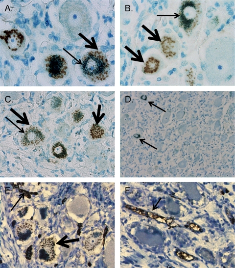
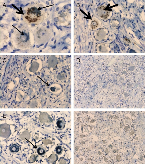
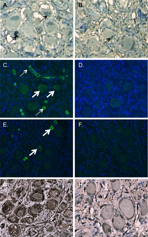
Analyses of varicella-zoster virus (VZV) protein expression during latency have been discordant, with rare to many positive neurons detected. We show that ascites-derived murine and rabbit antibodies specific for VZV proteins in vitro contain endogenous antibodies that react with human blood type A antigens in neurons. Apparent VZV neuronal staining and blood type A were strongly associated (by a χ² test, α = 0.0003). Adsorption of ascites-derived monoclonal antibodies or antiserum with type A erythrocytes or the use of in vitro-derived VZV monoclonal antibodies eliminated apparent VZV staining. Animal-derived antibodies must be screened for anti-blood type A reactivity to avoid misidentification of viral proteins in the neurons of the 30 to 40% of individuals who are blood type A.


Figures

Fig 1
Fig 1
Apparent immunostaining of VZV proteins in human cadaver DRG using mouse ascites-derived VZV monoclonal antibodies is associated with the presence of blood type A histo-blood group antigens (HBGAs) in neuronal Golgi zones. Immunohistochemical staining of tissue sections from subject 4 using 3,3-diaminobenzidine (DAB) chromogen to detect immunoreactivity (brown deposits, thick arrows) and azure B counterstain, which colors neuronal melanin (green deposits, thin arrows). (A to C) Magnification, ×400. Stained with a 1:100 dilution of MAB8612 (anti-gE) (A), MAB8616 (anti-IE62) (B), or MAB8614 (anti-ORF40) (C). (D) Stained with tissue culture-derived mouse Ig; arrows denote neuronal melanin. Magnification, ×100. (E and F) Staining of subject 4 (E) and subject 5 (F) with anti-A antibody at a 1:10 dilution. Thin arrows denote endothelial cell staining observed in all subjects with blood type A; the thick arrow denotes staining of blood type A antigens in the neuronal cytoplasm which were present in 8 of 10 subjects and indistinguishable from the signal observed when using the VZV monoclonal antibodies.
Fig 2
Fig 2
Apparent VZV immunoreactivity and neuronal anti-type A reactivity are associated with the mouse ascites Golgi (MAG) staining artifact. DRG tissue sections were stained with anti-MAG antibody (1:64,000 dilution), which was generated in mice by pristane priming followed by intraperitoneal injection of hybridoma cells that were not from immunized mice. The DAB antibody-specific signal is brown (thick arrows), and the melanin counterstain is green (thin arrows). Panels A and B are sections from subject 4, who is blood type A, subtype A1; panels C and D are sections from subject 5, who is blood type A, subtype A2; and panels E and F are sections from subject 1, who is blood type O. Two representative images are shown for each condition. Magnification for panels on the left side, ×400. Magnification for panels on the right side, ×200.
Fig 3
Fig 3
Elimination of apparent VZV immunoreactivity in neurons using tissue culture-derived VZV monoclonal antibodies or by adsorption using blood type A erythrocytes. (A and B) Immunohistochemical staining of tissue sections from a blood type A subject with purified hybridoma supernatants from anti-IE62 (A) and anti-gE (B) clones at a 1:10 dilution eliminates immunoreactivity. Sections are counterstained with azure B, which stains neuromelanin green (thin arrows). (C to F) Immunofluorescent staining using a rabbit polyclonal antibody to IE62 and Alexa Fluor 488-conjugated anti-rabbit secondary antibody (C and D) or gE mouse ascites-derived monoclonal antibody and Alexa Fluor 488-conjugated anti-mouse secondary antibody (E and F). The primary antibody was adsorbed with blood type A erythrocytes (D and F) or unadsorbed (C and E). Thin arrows denote endothelial staining, and thick arrows denote staining of neuronal histo-blood group A determinants in Golgi zones. Sections for immunofluorescent staining were pretreated with Sudan black to eliminate the signal from neuronal pigments and counterstained with DAPI (4′,6-diamidino-2-phenylindole). (G and H) Immunohistochemical staining of VZV-infected DRG (G) and uninfected DRG (H) from a SCID mouse-human xenograft model. A VZV-specific signal with gE-mouse ascites-derived monoclonal antibody after adsorption with blood type A erythrocytes demonstrates that adsorption of endogenous anti-type A antibodies does not alter VZV-specific immunoreactivity. Sections are counterstained with hematoxylin.

References

    1. Arend P. 2011. “Natural” antibodies and histo-blood groups in biological development with respect to histo-blood group A. Immunobiology 216:1318–1321 - PubMed
    1. Arend P, Nijssen J. 1977. A-specific autoantigenic ovarian glycolipids inducing production of ‘natural’ anti-A antibody. Nature 269:255–257 - PubMed
    1. Baumgarth N, Tung JW, Herzenberg LA. 2005. Inherent specificities in natural antibodies: a key to immune defense against pathogen invasion. Springer Semin. Immunopathol. 26:347–362 - PubMed
    1. Dodd J, Solter D, Jessell TM. 1984. Monoclonal antibodies against carbohydrate differentiation antigens identify subsets of primary sensory neurones. Nature 311:469–472 - PubMed
    1. Finstad CL, et al. 1991. Some monoclonal antibody reagents (C219 and JSB-1) to P-glycoprotein contain antibodies to blood group A carbohydrate determinants: a problem of quality control for immunohistochemical analysis. J. Histochem. Cytochem. 39:1603–1610 - PubMed

Publication types

MeSH terms