. 2012 Jan;86(1):69-80.
doi: 10.1128/JVI.06119-11. Epub 2011 Oct 19.

Mutations at the base of the icosahedral five-fold cylinders of minute virus of mice induce 3'-to-5' genome uncoating and critically impair entry functions

Affiliations

Mutations at the base of the icosahedral five-fold cylinders of minute virus of mice induce 3'-to-5' genome uncoating and critically impair entry functions

Susan F Cotmore et al. J Virol. 2012 Jan.

Abstract

The linear single-stranded DNA genome of minute virus of mice can be ejected, in a 3'-to-5' direction, via a cation-linked uncoating reaction that leaves the 5' end of the DNA firmly complexed with its otherwise intact protein capsid. Here we compare the phenotypes of four mutants, L172T, V40A, N149A, and N170A, which perturb the base of cylinders surrounding the icosahedral 5-fold axes of the virus, and show that these structures are strongly implicated in 3'-to-5' release. Although noninfectious at 37°C, all mutants were viable at 32°C, showed a temperature-sensitive cell entry defect, and, after proteolysis of externalized VP2 N termini, were unable to protect the VP1 domain, which is essential for bilayer penetration. Mutant virus yields from multiple-round infections were low and were characterized by the accumulation of virions containing subgenomic DNAs of specific sizes. In V40A, these derived exclusively from the 5' end of the genome, indicative of 3'-to-5' uncoating, while L172T, the most impaired mutant, had long subgenomic DNAs originating from both termini, suggesting additional packaging portal defects. Compared to the wild type, genome release in vitro following cation depletion was enhanced for all mutants, while only L172T released DNA, in both directions, without cation depletion following proteolysis at 37°C. Analysis of progeny from single-round infections showed that uncoating did not occur during virion assembly, release, or extraction. However, unlike the wild type, the V40A mutant extensively uncoated during cell entry, indicating that the V40-L172 interaction restrains an uncoating trigger mechanism within the endosomal compartment.


Figures
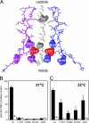
Fig 1
Fig 1
Mutations at the base of the 5-fold cylinder make MVMp virions temperature sensitive for growth. (A) Molecular model, made using Deep View Swiss PDB viewer and PDB 1MVM, depicting a cross-section of a single 5-fold cylinder. Amino acid residues from two of the five VP subunits that form the cylinder walls are represented as stick diagrams. Residues L172, N149, N170, and V40, which were mutated for this study, are highlighted as space-filling renditions. A space-filling rendition of a single glycine-rich VP2 sequence is modeled traversing the pore. This comprises a string of 10 glycine residues, between positions 28 and 39, interrupted by two serine residues at positions 30 and 35, as indicated. (B and C) Virus expansion assays in A9 cells transfected with wild-type (wt) or mutant MVMp-based infectious clones, cultured in the presence (black bars) or absence (white bars) of a neutralizing anticapsid antibody at either 37°C (B) or 32°C (C) and stained for NS1 on days 3 or 4 after transfection, respectively. Experiments were performed in duplicate, with each data point representing between 500 and 1,000 cells.
Fig 2
Fig 2
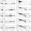
Gradient profiles of viruses generated in multiple-round infections. Iodixanol gradient analyses of wild-type and mutant virus stocks, as indicated, grown at 32°C through multiple cycles in A9 cells are shown. The left side of each pair of panels shows the distribution of capsid proteins in gradient fractions detected by Western transfer using the “allo-p” antipeptide antibody described in Materials and Methods. Lane 1 in each of these blots contained a 50-ng capsid protein standard. Iodixanol percentages in the original step gradient are indicated at the bottom of the figure, and the vertical dotted line between fractions 8 and 9 indicates a loading transition, with lanes representing fractions 2 to 8 each receiving 4-fold more sample than those representing fractions 9 to 13. The right side of each pair of panels shows nuclease-protected viral DNA associated with each fraction, detected following electrophoresis of equal aliquots of each sample through denaturing gels, Southern transfer, and hybridization with a full-length MVMp genomic probe. Positions of MVM restriction fragments used as molecular size markers are indicated, in kilobase pairs, on the right side of each blot.
Fig 3
Fig 3
Mapping the origin of subgenomic DNA found in multiple-round infections. (A) Samples from fractions 4 to 8 of the V40A gradient shown in Fig. 2E were digested with micrococcal nuclease and separated by electrophoresis though denaturing gels. Southern transfers after hybridization with 32P-labeled oligonucleotide probes specific for the negative strand at the positions indicated on a linear depiction of the MVM genome are shown. The positions of major bands between ∼3.5 and 2.3 kb, labeled a, b, and c, are diagrammed at the top. (B) Southern blots of fractions 6 to 10 from the L172T gradient in Fig. 2B, probed with the left-end or right-end probes shown in panel A.
Fig 4
Fig 4
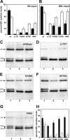
Ability of viruses to initiate infection before and after proteolysis. (A and B) Ability of wild-type and mutant viruses to initiate infection in A9 cells, as measured by the percentage of cells that express NS1. Cells were infected at 5,000 (A) or 50,000 (B) genomes/cell and cultured at 37°C (black bars) or 32°C (white bars). Experiments were performed in duplicate, with each data point representing between 500 and 1,000 cells. (C through G) Western blots of wild-type virions (C) and virions of mutant L172T (D), N149A (E), N170A (F), or V40A (G), either before trypsin digestion (lane 1) or after digestion at 37°C (lane 2) or room temperature (lane 3). Western blots were developed using a mixture of anti-allo-p-specific and VP1 N terminus-specific antipeptide antibodies, as described in Materials and Methods. (H) Samples from each of these reactions were then tested for their ability to initiate infection, which was quantitated by assessing the percentage of cells expressing NS1 after 26 h. Black bars represent untreated samples, while gray and white bars represent samples subjected to trypsin treatment at 37°C and room temperature, respectively.
Fig 5
Fig 5
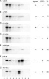
In vitro induction of conformational changes that expose L172T DNA. (A through F) Wild-type or L172T mutant virus samples that were incubated with or without trypsin and at various temperatures as shown and then analyzed for evidence of conformational changes by banding through analytical iodixanol gradients. Gradient fractions were subjected to electrophoresis through denaturing gels and analyzed by Southern blotting. Virions that had resisted conformational changes banded in fractions 2 to 4, while uncoated particles banded in fractions 6 to 8. Estimates of the fraction that was uncoated (labeled “%”) compare the DNA accumulation in fractions 6 to 8 with the total in fractions 2 to 4 and 6 to 8 combined. (G and H) Samples were treated in a similar way except that these virions were diluted in 3 volumes of TE8.7 buffer and stored at 4°C for 2 days prior to testing. This procedure chelates cations and primes the particles for subsequent heat-induced uncoating. (I through K) Southern transfers of gradient fractions from panel B (i.e., L172T digested with trypsin at 37°C) that were digested with micrococcal nuclease prior to electrophoresis. (I) The Southern blot was hybridized with a random-primed full-length genomic probe. (J and K) Nuclease-treated fractions 5 to 9 from the same gradient, hybridized with a random-primed probe representing either MVM nt 789 to 1043 (left-end probe) (J) or nt 3976 to 4154 (right-end probe) (K).
Fig 6
Fig 6
Induction of conformational changes in the V40A and N149A mutants. (A through D) Gradient analyses of V40A or N149A virions following exposure to 37°C, with or without trypsin as shown. Virions were analyzed for evidence of conformational changes by banding through analytical iodixanol gradients. Gradient fractions were subjected to electrophoresis through denaturing gels and analyzed by Southern blotting as described in the legend for Fig. 5. (E through G) Samples that were cation depleted prior to analysis by dilution in 3 volumes of TE8.7 and storage at 4°C for 4 h and analyzed as described above.
Fig 7
Fig 7
Viral DNA synthesis during single-round infections. (A and B) Total intracellular viral DNA generated in single-round infections at 37°C and 32°C, respectively, analyzed by Southern blotting after electrophoresis through nondenaturing gels. (C) Intracellular virion DNA in a TE8.7 extract of cells cultured at 32°C, following digestion with micrococcal nuclease and electrophoresis through a denaturing gel. (D) Virion DNA in samples of culture medium (containing neuraminidase) from the 32°C incubation, following nuclease digestion and analysis on a denaturing gel.
Fig 8
Fig 8
Mutant virions uncoat during cell entry. (A to C) Incoming virion DNA in total cell extracts following fractionation through iodixanol gradients and without in vitro nuclease treatment. Before infection cells were synchronized in G0 by isoleucine deprivation and then infected with wild-type or V40A virions at 32°C (A and B, respectively) or left uninfected (C) in complete medium plus aphidicolin to allow cell cycle progression to the G1-S border without entry into S phase. Neuraminidase was added at 24 h postinfection to release noninternalized virus, and cells were harvested 4 h later. Prior to cell lysis V40A virus equivalent to 1/10 of the experimental inoculum was added to the uninfected control cells (C), and these were then processed in parallel with test groups. This served as a control for any cleavage or modification that might occur during subsequent extraction and processing. (D) Fractions 4 to 9 of the V40A gradient (from panel B) digested with nuclease prior to electrophoresis and probed with ∼200-bp sequences from positions along the MVM genome, as indicated.

Similar articles

Cited by

References

    1. Agbandje-McKenna M, Llamas-Saiz AL, Wang F, Tattersall P, Rossmann MG. 1998. Functional implications of the structure of the murine parvovirus, minute virus of mice. Structure 6:1369–1381 - PubMed
    1. Bleker S, Sonntag F, Kleinschmidt JA. 2005. Mutational analysis of narrow pores at the fivefold symmetry axes of adeno-associated virus type 2 capsids reveals a dual role in genome packaging and activation of phospholipase A2 activity. J. Virol. 79:2528–2540 - PMC - PubMed
    1. Bubeck D, et al. 2005. The structure of the poliovirus 135S cell entry intermediate at 10-angstrom resolution reveals the location of an externalized polypeptide that binds to membranes. J. Virol. 79:7745–7755 - PMC - PubMed
    1. Carrasco C, et al. 2009. The capillarity of nanometric water menisci confined inside closed-geometry viral cages. Proc. Natl. Acad. Sci. U. S. A. 106:5475–5480 - PMC - PubMed
    1. Chandran K, Parker JS, Ehrlich M, Kirchhausen T, Nibert ML. 2003. The delta region of outer-capsid protein micro 1 undergoes conformational change and release from reovirus particles during cell entry. J. Virol. 77:13361–13375 - PMC - PubMed

Publication types

LinkOut - more resources