. 2011 Dec 16;286(50):43272-81.
doi: 10.1074/jbc.M111.290122. Epub 2011 Oct 19.

Structure function analysis of an ADP-ribosyltransferase type III effector and its RNA-binding target in plant immunity

Affiliations

Structure function analysis of an ADP-ribosyltransferase type III effector and its RNA-binding target in plant immunity

Byeong-ryool Jeong et al. J Biol Chem. .

Abstract

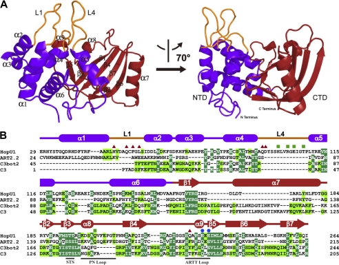
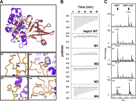
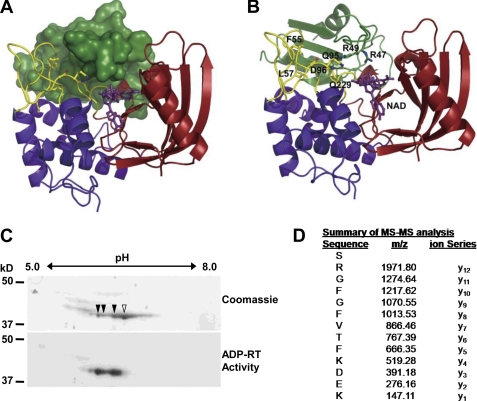
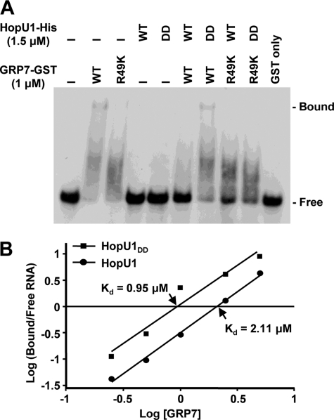
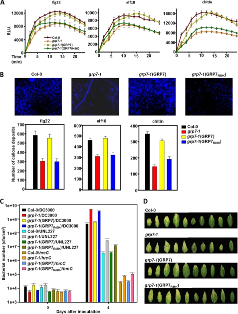
The Pseudomonas syringae type III effector HopU1 is a mono-ADP-ribosyltransferase that is injected into plant cells by the type III protein secretion system. Inside the plant cell it suppresses immunity by modifying RNA-binding proteins including the glycine-rich RNA-binding protein GRP7. The crystal structure of HopU1 at 2.7-Å resolution reveals two unique protruding loops, L1 and L4, not found in other mono-ADP-ribosyltransferases. Site-directed mutagenesis demonstrates that these loops are essential for substrate recognition and enzymatic activity. HopU1 ADP-ribosylates the conserved arginine 49 of GRP7, and this reduces the ability of GRP7 to bind RNA in vitro. In vivo, expression of GRP7 with Arg-49 replaced with lysine does not complement the reduced immune responses of the Arabidopsis thaliana grp7-1 mutant demonstrating the importance of this residue for GRP7 function. These data provide mechanistic details how HopU1 recognizes this novel type of substrate and highlights the role of GRP7 in plant immunity.


Figures

FIGURE 1.
FIGURE 1.
Structure of HopU1. A, overall structure of P. syringae pv. tomato DC3000 HopU1 in a ribbon representation. The N-terminal domain is colored in purple, C-terminal domain in red, and protruding loops L1 and L4 are shown in orange. Secondary structural elements are labeled, and two different views of the structure are shown. B, structure-based sequence alignment of HopU1 and three representative ADP-ribosyltransferases, rat ecto-ADP-ribosyltransferase ART2.2 (PDB code 1GXZ), ADP-ribosyltransferase C3bot2 from C. botulinum (PDB code 1R45), and C. limosum C3 exoenzyme (PDB code 3BW8). Identical residues are highlighted with dark green background, and highly conserved residues with light green background. Secondary structural elements are colored as in A and are shown above their corresponding sequences. Conserved motifs are indicated below their corresponding sequences. Residues that are critical for GRP7 recognition and enzymatic activity are indicated by red triangles, residues critical for enzymatic activity but not GRP7 recognition are indicated by blue dots, and those not critical for substrate recognition or activity are indicated by green squares.
FIGURE 2.
FIGURE 2.
Effect of HopU1 mutations on activity and GRP7 recognition. A, structure of HopU1 indicating the location of HopU1 mutations M1–M4 (detailed in bottom panels). The residues changed to Ala are highlighted and shown as stick representations. The structure was rotated ∼90° about the horizontal axis compared with the view presented on the left side of Fig. 1A. B, isothermal titration calorimetry of wild-type HopU1 and HopU1 derivatives M1–M4 with GRP7, indicating the extent that the HopU1 derivatives can interact with GRP7. C, electrospray ionization-mass spectrometry results of mADP-RT activity for wild-type HopU1 and HopU1 derivatives. The peak of substrates and products of each enzymatic reaction are indicated. The molecular mass of GRP7 (1–90 with 4 additional residues (Gly, Pro, His, and Met) left over after affinity tag cleavage) is 11,260 Da and GRP7 with an ADP-ribose modification is 11,800 Da. The mutations of each HopU1 mutant are as follows: M1, HopU1H49A/Q53A; M2, HopU1C54A/F55A/L57A; M3, HopU1Q95A/D96A; and M4, HopU1H100A/R103A/E105A/T108A.
FIGURE 3.
FIGURE 3.
Arginine 49 of GRP7 is ADP-ribosylated by HopU1. A, structure model of HopU1-NAD-GRP7-RRM complex. HopU1 is shown in ribbon representation, and the N-terminal domain is colored in blue, C-terminal domain in red, and protruding loops L1 and L4 are shown in yellow. GRP7-RRM (dark green) is shown in surface representation, and NAD (purple) is shown in stick representation. B, structure model with GRP7-RRM shown as ribbon representation. Critical HopU1 residues for interaction with GRP7 and potential ADP-ribosylation sites Arg-47 and Arg-49 are indicated in stick representation. C, two-dimensional PAGE gels of in vitro mADP-RT reactions containing purified HopU1-His, GRP7-GST, and 32P-labeled NAD stained with Coomassie Blue to visualize total protein or exposed to autoradiography film to identify 32P-labeled proteins. Protein spots labeled with 32P corresponding to ADP-ribosylated proteins are marked with filled arrowheads, and the unlabeled spot is marked with an open arrowhead. D, mass spectrometric analyses of tryptic peptides derived from the above mADP-RT reactions using nonradioactive NAD. All spots corresponding to the indicated spots in B were cut out and sent for MS/MS. One fragment shown contained a higher molecular mass than predicted. The molecular mass of the arginine (y12) corresponds to Arg-49 of GRP7 and was equal to ADP-ribosylated arginine, indicating that this residue was ADP-ribosylated by HopU1.
FIGURE 4.
FIGURE 4.
ADP-ribosylation by HopU1 reduces ability of GRP7 to bind RNA. A, electrophoretic mobility shift assay of an RNA probe with GRP7-GST after treatment with HopU1-His or its catalytic inactive mutant HopU1DD-His. Standard ADP-ribosylation reactions were performed with GRP7-GST or GRP7-GSTR49K in the presence of HopU1 or HopU1DD, and a 32P-labeled probe (ATGRP7 UTR WT) was added to each reaction mix. These were run on native polyacrylamide gels and exposed to x-ray films. B, similar assays done with differing amounts of GRP7-GST. The protein-bound and free RNA probes were quantified using a PhosphorImager scanner. The ratio of protein-bound and free forms of RNA was plotted against the concentration of GRP7 using a logarithmic scale. The x intercept allowed estimation of Kd for both HopU1 and HopU1DD as indicated. The autoradiogram used to calculate the kDa is shown in supplemental Fig. S4.
FIGURE 5.
FIGURE 5.
An Arabidopsis grp7 mutant is defective in PTI responses and more susceptible to P. syringae, and these phenotypes are complemented by wild-type GRP7 but not by a GRP7 derivative that is reduced in its ability to bind to RNA. A and B, ROS levels in relative light units (RLU) (A) and callose (B) in wild-type Arabidopsis Columbia-0 (Col-0), the Columbia-0 grp7-1 mutant, the grp7-1 mutant complemented with wild-type GRP7 (grp7-1(GRP7)), and the grp7-1 mutant complemented with a GRP7 derivative containing a lysine instead of an arginine at position 49 (grp7-1(GRP7R49K)) after treatment with flg22, elf18, or chitin. C, bacterial growth in the plants listed in A of wild-type P. syringae pv. tomato DC3000, a DC3000 polyeffector mutant lacking about one third of its type III effector inventory (UNL227), and a DC3000 mutant with a defective T3SS (hrcC). D, disease symptoms at day 4 on Arabidiopsis plants described in A after infection with wild-type DC3000.

References

    1. Büttner D., He S. Y. (2009) Plant Physiol. 150, 1656–1664 - PMC - PubMed
    1. Alfano J. R., Collmer A. (2004) Annu. Rev. Phytopathol. 42, 385–414 - PubMed
    1. Hann D. R., Gimenez-Ibanez S., Rathjen J. P. (2010) Curr. Opin. Plant Biol. 13, 388–393 - PubMed
    1. Block A., Alfano J. R. (2011) Curr. Opin. Microbiol. 14, 39–46 - PMC - PubMed
    1. Boller T., Felix G. (2009) Annu. Rev. Plant Biol. 60, 379–406 - PubMed

Publication types

MeSH terms

Associated data