. 2011 Dec 9;286(49):42232-42247.
doi: 10.1074/jbc.M111.262295. Epub 2011 Oct 19.

Curcumin enhances the efficacy of chemotherapy by tailoring p65NFκB-p300 cross-talk in favor of p53-p300 in breast cancer

Affiliations

Curcumin enhances the efficacy of chemotherapy by tailoring p65NFκB-p300 cross-talk in favor of p53-p300 in breast cancer

Gouri Sankar Sen et al. J Biol Chem. .

Abstract

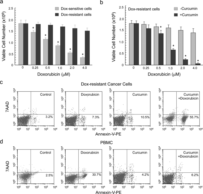
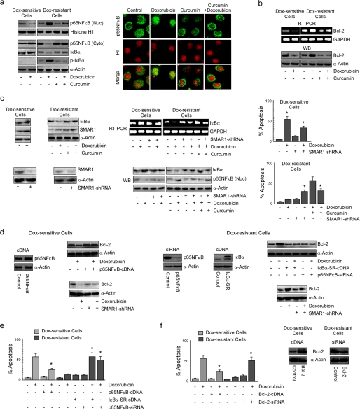
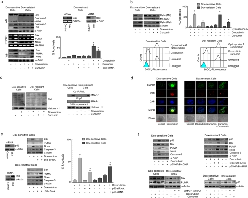
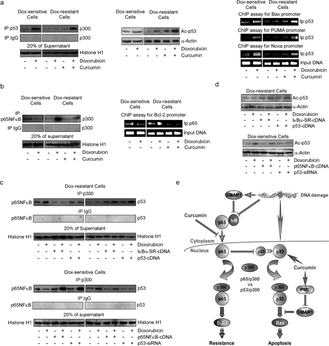
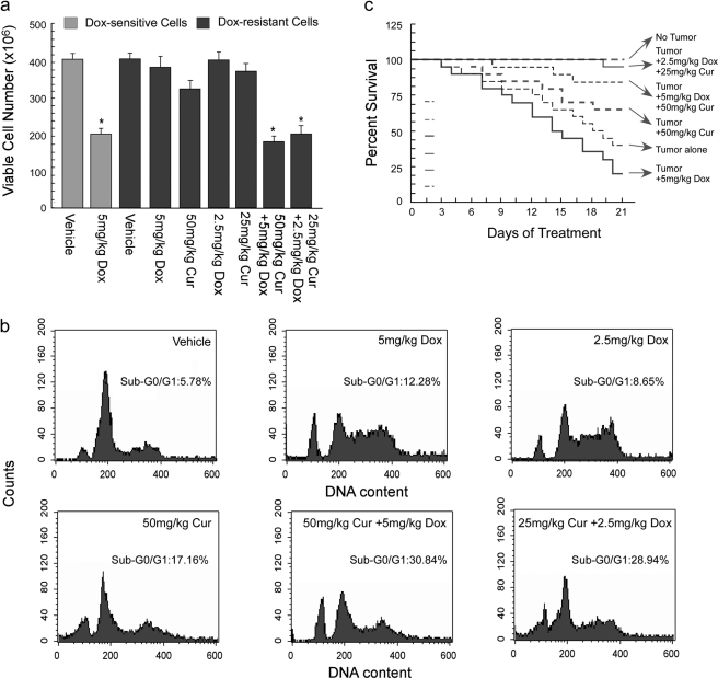
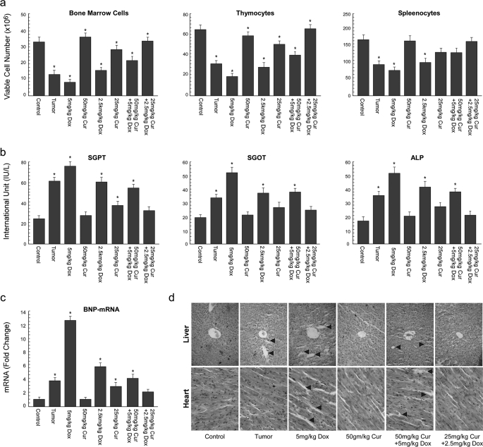
Breast cancer cells often develop multiple mechanisms of drug resistance during tumor progression, which is the major reason for the failure of breast cancer therapy. High constitutive activation of NFκB has been found in different cancers, creating an environment conducive for chemotherapeutic resistance. Here we report that doxorubicin-induced SMAR1-dependent transcriptional repression and SMAR1-independent degradation of IkBα resulted in nuclear translocation of p65NFκB and its association with p300 histone acetylase and subsequent transcription of Bcl-2 to impart protective response in drug-resistant cells. Consistently SMAR1-silenced drug-resistant cells exhibited IkBα-mediated inhibition of p65NFκB and induction of p53-dependent apoptosis. Interestingly, curcumin pretreatment of drug-resistant cells alleviated SMAR1-mediated p65NFκB activation and hence restored doxorubicin sensitivity. Under such anti-survival condition, induction of p53-p300 cross-talk enhanced the transcriptional activity of p53 and intrinsic death cascade. Importantly, promyelocyte leukemia-mediated SMAR1 sequestration that relieved the repression of apoptosis-inducing genes was indispensable for such chemo-sensitizing ability of curcumin. A simultaneous decrease in drug-induced systemic toxicity by curcumin might also have enhanced the efficacy of doxorubicin by improving the intrinsic defense machineries of the tumor-bearer. Overall, the findings of this preclinical study clearly demonstrate the effectiveness of curcumin to combat doxorubicin-resistance. We, therefore, suggest curcumin as a potent chemo-sensitizer to improve the therapeutic index of this widely used anti-cancer drug. Taken together, these results suggest that curcumin can be developed into an adjuvant chemotherapeutic drug.


Figures

FIGURE 1.
FIGURE 1.
Treatment of drug-resistant ascites carcinoma cells with curcumin restored their sensitivity toward doxorubicin. a, 1 million doxorubicin-sensitive and 1 million doxorubicin-resistant ascites carcinoma cells were treated with a dose range (0–4 μm) of doxorubicin for a time interval of 24 h and subjected to determination of cell viability test by trypan blue dye exclusion assay. b, to assay the in vitro chemosensitizing potential of curcumin, doxorubicin-resistant cells were pretreated with 10 μm curcumin for a period of 2 h followed by 24 h of doxorubicin treatment. Doxorubicin-resistant cells (c) and peripheral blood mononuclear cells (PBMC; d) were treated with doxorubicin (1 μm) and curcumin (10 μm) separately or in combination and subjected to flow cytometric determination of percentage apoptosis by annexin V-PE/7AAD binding. Values are the mean ± S.E. of five independent experiments or are representative of a typical experiment in the case of flow cytometric analysis. *. p < 0.05 when compared with respective control sets.
FIGURE 2.
FIGURE 2.
Curcumin inhibits drug-induced NFκB activation and Bcl-2 up-regulation maneuvering SMAR1 to induce apoptosis of doxorubicin-resistant cancer cells. a, doxorubicin-treated drug-sensitive cells and doxorubicin/curcumin alone or in combination-treated drug-resistant cells were subjected to isolation of nuclear and cytosolic fractions for Western blot (WB) analysis or to confocal microscopy and evaluated for nuclear localization of p65NFκB after 1 h of doxorubicin treatment. At the same time cell lysates from the same experimental set up was verified for IκBα and phospho-IκBα levels. The concentration of doxorubicin and curcumin were 1 and 10 μm, respectively. Magnification bars in confocal microscopy images indicate 20 μm. b, RT-PCR and Western blot images depict expression of Bcl-2 at 24 h in the same set of experiments. c, at the same time the lysates from the previous experimental sets were screened for IκBα and SMAR1 levels. To study the role of SMAR1 in regulation of IκBα expression, SMAR1-shRNA-transfected drug-sensitive or drug-resistant cells were treated with doxorubicin or in combination with curcumin to follow the expression of IκBα by RT-PCR and Western blot analysis. In parallel, a portion of the same set of cells was subjected to Western blot analysis to determine nuclear translocation of p65NFκB, and the remaining portion was screened for percentage apoptosis. d, p65NFκB-cDNA-/SMAR1-shRNA-transfected drug-sensitive and IκBα-SR-cDNA-/p65NFκB-siRNA-/SMAR1-shRNA-transfected drug-resistant cells were treated with doxorubicin for 24 h and explored for Bcl-2 expression by Western blot analysis. e, flow cytometric determination of doxorubicin-induced apoptosis in p65NFκB-cDNA-transfected drug-sensitive and IκBα-SR-cDNA-/p65NFκB-siRNA-transfected drug-resistant cells is shown. f, similarly, Bcl-2-cDNA-transfected drug-sensitive cells and Bcl-2-siRNA-transfected drug-resistant cells were evaluated for doxorubicin sensitivity by measuring annexin V-PE/7AAD positivity. GAPDH and α-actin/histone H1 were used as internal loading controls for Western blot and RT-PCR. Values are the mean ± S.E. of five independent experiments in each case or are representative of a typical experiment in the case of RT-PCR analysis and Western blots. Confocal images shown are representative of >10 images taken in different fields from two independent experiments. *, p < 0.05 when compared with respective untransfected/control sets.
FIGURE 3.
FIGURE 3.
Curcumin-mediated NFκB inhibition triggers p53-dependent Bax activation in doxorubicin-resistant cancer cells. a, sensitive/resistant cells were treated with doxorubicin alone or in combination with curcumin for 24 h and Western-blotted (WB) for the evaluation of (pro)active-caspase-9 and caspase-3. In parallel, p53 levels in the nuclear fractions was also determined. Expression levels of Bax, PUMA, and Noxa in the same experimental set were determined by RT-PCR and Western blot analysis. Control/Bax-siRNA-transfected resistant cells were treated with doxorubicin alone or in combination with curcumin, and percent apoptosis was determined by annexin V-PE/7AAD positivity. b, sensitive/resistant cells were treated with doxorubicin alone or in combination with curcumin and were subjected to isolation of cytosolic and mitochondrial fractions and Western-blotted for cytochrome c (Cyt c). In parallel, flow cytometry was used to determine mitochondrial transmembrane potential and percentage apoptosis in doxorubicin/combination dose-treated sensitive/resistant cells in the presence and absence of cyclosporine A. Mn-SOD, manganese superoxide dismutase antibody. c, lysates of sensitive/resistant cells treated with doxorubicin alone or in combination with curcumin for 24 h were Western-blotted with anti-PML antibody or immunoprecipitated with PML/IgG, and the immunoprecipitates (co-IP) were Western-blotted with SMAR1. d, confocal images depict co-localization of SMAR1 with PML in sensitive/resistant cells treated with doxorubicin alone or in combination with curcumin, respectively. Bar length in confocal microscopy images indicate 10 μm (e) p53-siRNA-transfected sensitive cells, and p53-cDNA-transfected resistant cells were treated with doxorubicin and evaluated for Bax, PUMA, Noxa, and caspase-3 levels at 24 h by Western blot and percent apoptosis by flow cytometry. f, IκBα-SR-cDNA-/p65NFκB-siRNA-transfected resistant cells and p65NFκB-overexpressed sensitive cells were evaluated for change in p53, Bax, PUMA, Noxa, and caspase-3 levels at 24 h. SMAR1-shRNA-transfected sensitive/resistant cells were treated with doxorubicin alone or in combination with curcumin, respectively, and the expression of p53 at 8 h or Bax and PUMA at 24 h was assayed by Western blot analysis. GAPDH and α-actin were used as internal loading controls for RT-PCR and Western blot. Values are the mean ± S.E. of five independent experiments in each case or is representative of a typical experiment in the case of RT-PCR and Western blot analysis. Confocal images shown are representative of >10 images taken in different fields from two independent experiments. *, p < 0.05 when compared with respective untransfected/control sets.
FIGURE 4.
FIGURE 4.
Curcumin induced p53-p300 interaction by inhibiting drug-induced NFκB activation in doxorubicin-resistant cells. a, left panel; b, left panel, p53/p65NFκB-associated p300 was immunopurified with anti-p53/p65NFκB antibodies from nuclear lysates of sensitive/resistant cells treated with doxorubicin alone or in combination with curcumin for 24 h and were Western-blotted (WB) with p300 antibody. A portion of nuclear lysates from the same set were used for Western blot analysis of acetylated p53 at lysine 373 (a, middle panel), and remaining cells from the same experimental set were subjected to ChIP assay for the determination of p53 and p65NFκB activity on Bax/PUMA/Noxa and Bcl-2 promoter, respectively (a, right panel; b, right panel). p53-cDNA/IκBα-SR-cDNA-transfected doxorubicin-resistant cells and p53-siRNA/p65NFκB-cDNA-transfected doxorubicin-sensitive cells were treated with doxorubicin for 24 h, and the nuclear lysates obtained were either subjected to immunoprecipitation (IP) with p300/IgG antibody and the immunoprecipitates were Western-blotted with anti-p65NFκB/-p53 antibodies (c) or were subjected to Western blot analysis of acetylated p53 at lysine 373 (d). To verify comparable protein input during immunoprecipitation, 20% of supernatant from the nuclear lysates was blotted with histone H1 antibody. Values are the mean ± S.E. of five independent experiments in each case or representative of typical experiment in case of ChIP assay and Western blots. *, p < 0.05 when compared with respective untransfected/control sets. e, shown is a schematic illustration depicting differential regulation of anti- and pro-apoptotic network by curcumin in drug-resistant cells.
FIGURE 5.
FIGURE 5.
Curcumin reverted drug resistance and provided survival advantage of doxorubicin-treated tumor-bearing mice. a, doxorubicin-sensitive/resistant tumor-bearing mice were treated with doxorubicin alone or in combination with curcumin at different doses, and the total number of viable tumor cells in the peritoneal cavity was assayed by trypan blue dye-exclusion test. b, doxorubicin-resistant tumor-bearing mice were treated with doxorubicin alone or in combination with curcumin, and the tumor cells were subjected to flow cytometric determination of percentage hypoploidy (sub-G0/G1 phase cells). c, the survival rates of doxorubicin-resistant tumor-bearing animals treated with doxorubicin alone or in combination with curcumin were calculated by counting the number of live animals at a fixed time interval and were represented as Kaplan-Meier curve. Values are the mean ± S.E. of five independent experiments in each case. *, p < 0.05 when compared with respective control sets.
FIGURE 6.
FIGURE 6.
Curcumin ameliorated doxorubicin-induced systemic toxicity in drug-resistant tumor-bearing mice. a, total number of viable cells in the thymus, bone marrow, and spleen from normal and doxorubicin/curcumin (Cur) alone or in combination-treated doxorubicin-resistant tumor-bearing mice were determined by trypan blue dye-exclusion assay. b, serum glutamate pyruvate transaminase (SGPT), glutamate oxaloacetate transaminase (SGOT), and alkaline phosphatase (ALP) from the above set of mice were assayed as described under “Experimental Procedures” and plotted graphically. c, real-time PCR experiments were carried out to compare -fold change in BNP-mRNA levels from the same set of mice and were plotted graphically (left panels). Histological sections of liver and heart from the animals were stained with hematoxylin and counter-stained with eosin and microscopically analyzed for histopathological examinations of tissue toxicity like cellular damage and vacuolization. Values are the mean ± S.E. of five independent experiments in each case. *, p < 0.05 when compared with respective control sets or representative of typical experiment in case histological analysis.

Similar articles

Cited by

References

    1. Chintamani, Tandon M., Mishra A., Agarwal U., Saxena S. (2011) World J. Surg. Oncol 9, 19. - PMC - PubMed
    1. Campbell K. J., O'Shea J. M., Perkins N. D. (2006) BMC Cancer 6, 101. - PMC - PubMed
    1. Arafa el-S. A., Zhu Q., Shah Z. I., Wani G., Barakat B. M., Racoma I., El-Mahdy M. A., Wani A. A. (2011) Mutat. Res. 706, 28–35 - PMC - PubMed
    1. Liu X., Wang B., Ma X., Guo Y. (2009) Jpn. J. Clin. Oncol. 39, 418–424 - PubMed
    1. Gionet N., Jansson D., Mader S., Pratt M. A. (2009) J. Cell. Biochem. 107, 448–459 - PubMed

Publication types

MeSH terms