. 2011 Dec 9;286(49):42325-42336.
doi: 10.1074/jbc.M110.210336. Epub 2011 Oct 19.

Characterization of the Src/Abl hybrid kinase SmTK6 of Schistosoma mansoni

Affiliations

Characterization of the Src/Abl hybrid kinase SmTK6 of Schistosoma mansoni

Svenja Beckmann et al. J Biol Chem. .

Abstract

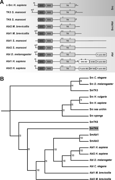
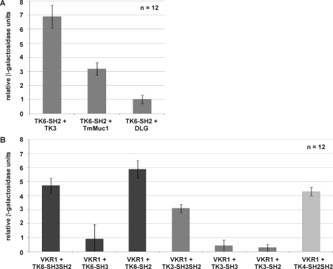
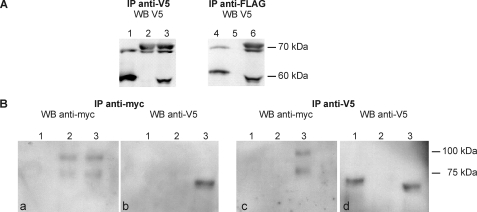
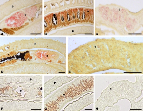
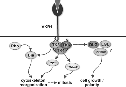
Cellular protein-tyrosine kinases play key roles in signal transduction processes in eukaryotes. SmTK4 was the first Syk kinase identified in a parasite and found to be tissue-specifically transcribed in the gonads of adult Schistosoma mansoni. Functional analyses confirmed its role in oogenesis and spermatogenesis. As an SmTK4 upstream binding partner, the cellular protein-tyrosine kinase SmTK6 was isolated from a yeast two-hybrid library. Phylogenetic analyses performed in this study confirmed the first suggestions of a hybrid character of SmTK6. Biochemical studies made in Xenopus oocytes using inhibitors against Src (herbimycin A) and Abl (imatinib) kinases exhibited a biochemical inhibition profile of SmTK6, which was intermediate of Src and Abl kinases. As SmTK6 upstream interaction partners, we identified among others the known Src kinase SmTK3 and the Venus kinase receptor SmVKR1 of S. mansoni by yeast two-hybrid analyses, all of which co-localized in the gonads. Co-immunoprecipitation experiments confirmed interactions between SmTK6 and SmTK3 or SmVKR1. In Xenopus oocytes, it was finally shown that SmVKR1 but also SmTK3 were able to activate SmTK6 enzymatic activity indicating its functions in a receptor tyrosine kinase signal transduction cascade. These results not only demonstrate an intermediate but Src-biased profile of the unusual kinase SmTK6. They also strongly substantiate previous indications for a kinase complex, consisting of a receptor tyrosine kinase, Syk and Src kinases, which has been hypothesized to be involved in proliferation and differentiation processes in the gonads of schistosomes.


Figures

FIGURE 1.
FIGURE 1.
A, schematic structures of the functional domains of c-Src from human, the Src kinase SmTK3 and the Src/Abl hybrid kinase SmTK6 from S. mansoni, Abl1 and Abl2 from M. brevicollis, Abl1 and Abl2 from S. mansoni, Abl from Drosophila melanogaster, and Abl1 and Abl2 from human. m, myristoylation site (m?, predicted but not verified myristoylation site); TK, tyrosine kinase domain; G BD, G-actin-binding domain; MT BD, microtubule-binding domain; F-actin BD, F-actin-binding domain; Y, conserved tyrosine phosphorylation site; gray triangle, nuclear location site (NLS). B, dendrogram of the phylogenetic analysis of the SH3-SH2-TK cassette sequences of the Src/Abl hybrid kinase SmTK6, the Src kinases SmTK3 and SmTK5, and the Abl kinases SmAbl1 and SmAbl2 of S. mansoni, as well as other metazoan CTKs using ClustalX and TreeViewX. Bootstrap values are indicated. Sequences were obtained from the National Center for Biotechnology Information using the Entrez Browser (www.ncbi.nlm.nih.gov) and from the M. brevicollis genome website. The corresponding protein accession numbers are as follows: Abl D. melanogaster (protein-tyrosine kinase Abl, D. melanogaster; AAA28934); Abl1 H. sapiens (tyrosine-protein kinase Abl1 isoform b, H. sapiens; NP_009297); Abl2 H. sapiens (tyrosine-protein kinase Abl2 isoform b, H. sapiens; NP_009298); Abl C. elegans (tyrosine-protein kinase Abl-1, C. elegans; P03949); SmAbl1 (Abl protein-tyrosine kinase 1, S. mansoni; CBH50761); SmAbl2 (Abl protein-tyrosine kinase 2, Schistosoma mansoni; CBH50762); Abl1 M. brevicollis (Abl protein kinase 1, M. brevicollis; XP_001742753); Abl2 M. brevicollis (Abl protein kinase 2, M. brevicollis; XP_001746037); Src C. elegans (protein-tyrosine kinase F49B2.5, C. elegans; CAB04427); Src D. melanogaster (Dsrc41, D. melanogaster; BAA07705); Src sponge (tyrosine-protein kinase isoform SRK1, Spongilla lacustris; P42686); Src human (proto-oncogene tyrosine-protein kinase SRC, H. sapiens; NP_005408); Src sea urchin (Src-type protein-tyrosine kinase, Anthocidaris crassispina; BAA33741); Src Hydra (Src-related protein-tyrosine kinase, Hydra vulgaris; AAA29217); SmTK5 (Src/Fyn tyrosine kinase 5, S. mansoni; AAF64151); SmTK3 (Src tyrosine kinase, S. mansoni; CAE51198); and SmTK6 (Src tyrosine kinase, S. mansoni; CAZ50862).
FIGURE 2.
FIGURE 2.
A, yeast cells (strain AH109) were re-transformed with one representative prey clone of each group together with the bait SmTK6-SH2 pBridge, and the relative β-gal activity was measured. Tested clones were (from left to right) as follows: SmTK3 (Src kinase), the mucin homolog SmTmMuc1, and the Discs large homolog SmDiscs large. The statistical evaluation of 12 independent measurements of β-gal activity (nbiol = 2, each with ntech = 6) is shown (error bars are indicated). B, yeast cells (strain AH109) were re-transformed individually with the prey plasmid SmVKR1-C-term pACT2 together with the baits (from left to right) SmTK6-SH3SH2 pBridge, SmTK6-SH3 pBridge, SmTK6-SH2 pBridge, SmTK3-SH3SH2 pBridge, SmTK3-SH3 pBridge, SmTK3-SH2 pBridge, or SmTK4-SH2SH2 pBridge. The relative β-gal activity was measured. The statistical evaluation of six independent measurements (nbiol = 2, each with ntech = 6) is shown (error bars are indicated). Dark gray columns represent the involvement of SmTK6 constructs; gray columns represent SmTK3 constructs, and the light gray column represents the SmTK4 construct.
FIGURE 3.
FIGURE 3.
A, tagged full-length variants of SmTK6 and SmTK3 were expressed alone or together in Xenopus oocytes (lanes 1 and 4, FLAG/V5 SmTK6-fl; lanes 2 and 5, V5-SmTK3-fl; lanes 3 and 6, FLAG/V5-SmTK6-fl + V5-SmTK3-fl), immunoprecipitated (IP) by anti-V5 (left) or anti-FLAG antibodies (right), and then analyzed by Western blot (WB) using anti-V5 antibodies. SmTK6 has been detected as expected at 55 kDa and SmTK3 with the expected size of 70 kDa. B, V5-SmTK6-fl and Myc-tagged SmVKR1YYRE-C-term were expressed alone or together in Xenopus oocytes (lane 1, V5-SmTK6-fl; lane 2, myc-SmVKR1YYRE-C-term; lane 3, V5-SmTK6-fl + myc-SmVKR1YYRE-C-term) imunoprecipitated (IP) by anti-Myc (left pair) or anti-V5 (right pair) antibodies, and then analyzed by Western blot using the same antibodies. SmTK6 is expected at 55 kDa and SmVKR1-C-term at 70 kDa (the additional upper band is supposed to represent post-translational modifications of VKR1).
FIGURE 4.
FIGURE 4.
Nonconserved regions of SmDLG (A–C) and SmVKR1 (D and E) served as templates to synthesize digoxigenin-labeled antisense transcripts used as probes for in situ hybridization. SmVKR1 signals were exclusively detected in the ovary (o), whereas SmDLG signals were detected in the ovary and the vitellarium (v) of the female as well as in the testes (t) of the male. p, parenchyma. For control, a digoxigenin-labeled sense probe was used (F–H). (Scale bar, 50 μm.)
FIGURE 5.
FIGURE 5.
In S. mansoni the CTKs SmTK3, SmTK4, and SmTK6 may be members of a trimeric complex, which interacts with the RTK SmVKR1. Results of a previous study had already indicated that SmTK3 also interacted with the diaphanous homolog SmDia, which is a binding partner of the Rho-GTPase SmRho1. Both SmDia and SmRho1 were suggested to be part of cooperative RTK and G-protein-coupled receptor signaling pathways integrating at SmDia to organize the actin cytoskeleton within the gonads of schistosomes (20). As downstream partners of SmTK4, MAPK-activating protein (PM20/21) and mapmodulin were found, which may be involved in cytoskeleton reorganization and mitosis (13). SmDLG as a binding partner of SmTK6 may become activated upon complex formation and may subsequently interact with SmLGL and Scribble to control processes of cell growth and/or cell polarity.

Similar articles

Cited by

References

    1. Hunter T. (2009) Curr. Opin. Cell Biol. 21, 140–146 - PMC - PubMed
    1. Bromann P. A., Korkaya H., Courtneidge S. A. (2004) Oncogene 23, 7957–7968 - PubMed
    1. Lemmon M. A., Schlessinger J. (2010) Cell 141, 1117–1134 - PMC - PubMed
    1. Takahashi A., Obata Y., Fukumoto Y., Nakayama Y., Kasahara K., Kuga T., Higashiyama Y., Saito T., Yokoyama K. K., Yamaguchi N. (2009) Exp. Cell Res. 315, 1117–1141 - PubMed
    1. Hubbard S. R., Till J. H. (2000) Annu. Rev. Biochem. 69, 373–398 - PubMed

Publication types

MeSH terms