. 2011 Dec;22(24):4908-17.
doi: 10.1091/mbc.E11-07-0614. Epub 2011 Oct 19.

Sequestration of phosphoinositides by mutated MARCKS effector domain inhibits stimulated Ca(2+) mobilization and degranulation in mast cells

Affiliations

Sequestration of phosphoinositides by mutated MARCKS effector domain inhibits stimulated Ca(2+) mobilization and degranulation in mast cells

Deepti Gadi et al. Mol Biol Cell. 2011 Dec.

Abstract

Protein kinase C β (PKCβ) participates in antigen-stimulated mast cell degranulation mediated by the high-affinity receptor for immunoglobulin E, FcεRI, but the molecular basis is unclear. We investigated the hypothesis that the polybasic effector domain (ED) of the abundant intracellular substrate for protein kinase C known as myristoylated alanine-rich protein kinase C substrate (MARCKS) sequesters phosphoinositides at the inner leaflet of the plasma membrane until MARCKS dissociates after phosphorylation by activated PKC. Real-time fluorescence imaging confirms synchronization between stimulated oscillations of intracellular Ca(2+) concentrations and oscillatory association of PKCβ-enhanced green fluorescent protein with the plasma membrane. Similarly, MARCKS-ED tagged with monomeric red fluorescent protein undergoes antigen-stimulated oscillatory dissociation and rebinding to the plasma membrane with a time course that is synchronized with reversible plasma membrane association of PKCβ. We find that MARCKS-ED dissociation is prevented by mutation of four serine residues that are potential sites of phosphorylation by PKC. Cells expressing this mutated MARCKS-ED SA4 show delayed onset of antigen-stimulated Ca(2+) mobilization and substantial inhibition of granule exocytosis. Stimulation of degranulation by thapsigargin, which bypasses inositol 1,4,5-trisphosphate production, is also substantially reduced in the presence of MARCKS-ED SA4, but store-operated Ca(2+) entry is not inhibited. These results show the capacity of MARCKS-ED to regulate granule exocytosis in a PKC-dependent manner, consistent with regulated sequestration of phosphoinositides that mediate granule fusion at the plasma membrane.


Figures

FIGURE 1:
FIGURE 1:
Dynamics of stimulated PKCβI-EGFP redistributions in IgE-sensitized RBL cells. (A) Time course of PKCβI-EGFP redistributions (green) in cells upon stimulation with antigen at 37°C (still frames from Supplemental Movie S1). Times in images specify intervals after antigen addition. Plot shows relative cytoplasmic fluorescence in the region of interest marked in a representative individual cell. Scale bar, 10 μm. (B) Time course of PKCβI-EGFP recruitment to the plasma membrane upon antigen stimulation at 25°C monitored as depletion of cytoplasmic fluorescence in a single representative cell. (C) Time course of PKCβI-EGFP recruitment to the plasma membrane upon stimulation with thapsigargin at 37°C monitored as depletion of cytoplasmic fluorescence for an average of six cells. (D) Time courses of PKCβI-EGFP recruitment to the plasma membrane monitored as depletion of cytoplasmic fluorescence (blue) and cytoplasmic [Ca2+] changes monitored by Fura Red (red) in a single representative cell stimulated with antigen at 37°C.
FIGURE 2:
FIGURE 2:
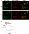
Live-cell imaging of antigen-stimulated mRFP-MARCKS-ED and PKCβI-EGFP oscillations. (A) Representative images showing the distributions of PKCβI-EGFP (green) and mRFP-MARCKS-ED (red) before (top) and 100 s after antigen addition at 25°C (bottom) (still frames from Supplemental Movies S2a and S2b). Scale bar, 10 μm. (B) Time courses of cytoplasmic changes of PKCβI-EGFP (blue) and mRFP-MARCKS-ED (red) in a region of interest indicated in representative RBL cell in A.
FIGURE 3:
FIGURE 3:
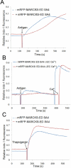
Association of mRFP-MARCKS-ED SA4 with the plasma membrane during stimulation by antigen. (A) Images of RBL cells transiently transfected with mutant mRFP-MARCKS-ED (red) and labeled with the [Ca2+] indicator Fluo-4 (green) at various times of stimulation by antigen at 25°C. Images show distributions of labels at time points before and after addition of antigen. Scale bar, 10 μm. (B) Fluorescence intensities from a cytoplasmic region of interest in a representative cell show that mRFP-MARCKS-ED SA4 (red) remains concentrated at the plasma membrane while stimulated [Ca2+] oscillations are observed. (C) Ca2+ mobilization in response to Ag at 25°C is delayed in a cell expressing mutant mRFP-MARCKS-ED (black) compared with an untransfected cell (blue); cells shown in Supplemental Movie S3. (D) Ca2+ response to antigen in the absence of extracellular Ca2+ is substantially delayed in a cell transiently expressing mutant mRFP-MARCKS-ED (red) compared with the response in an untransfected cell (blue) in the same sample.
FIGURE 4:
FIGURE 4:
Antigen-stimulated Ca2+ mobilization is delayed in cells stably expressing mRFP-MARCKS-ED SA4. (A) Steady-state fluorimetry responses to antigen in control RBL cells (blue) and cells stably expressing mRFP-MARCKS-ED SA4 (red). (B) Ca2+ response to antigen in the absence of extracellular Ca2+ is substantially delayed in RBL cells stably expressing mutant mRFP-MARCKS-ED (red) compared with the response in untransfected cells (blue). Subsequent addition of 1.8 mM [Ca2+] results in similar Ca2+ entry for each. (C) Ca2+ mobilization in response to thapsigargin in RBL cells stably expressing mRFP-MARCKS-ED SA4 (red) is similar compared with the response in untransfected RBL cells (blue).
FIGURE 5:
FIGURE 5:
MARCKS-ED SA4 inhibits antigen-stimulated degranulation monitored by real-time imaging. Degranulation monitored as FITC-dextran bursts from antigen-stimulated RBL cells shows delayed and reduced responses in cells expressing mRFP-MARCKS-ED SA4. Times in images indicate intervals after antigen addition; white arrow identifies a cell expressing mRFP-MARCKS-ED SA4. Scale bar, 10 μm. See Supplemental Movie S4.
FIGURE 6:
FIGURE 6:
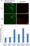
Inhibition of degranulation due to expression of MARCKS-ED SA4 as monitored by CD63 appearance at the plasma membrane. (A) Representative images showing reduced plasma membrane labeling by anti-CD63 due to antigen stimulation in cells expressing mutated MARCKS-ED expression. Scale bars, 10 μm. (B) Quantification of anti-CD63 labeling at the plasma membrane for unstimulated, antigen-stimulated, and thapsigargin-stimulated cells with and without expression of MARCKS-ED SA4. Error bars, SEM for 90–100 cells for each condition from n = 6 experiments. The p values from Student's t-test show **p < 0.01 for stimulated cells expressing MARCKS-ED SA4 compared with stimulated control cells.

Similar articles

Cited by

References

    1. Aderem A. The MARCKS brothers: a family of protein kinase C substrates. Cell. 1992;71:713–716. - PubMed
    1. Bai J, Tucker WC, Chapman ER. PIP2 increases the speed of response of synaptotagmin and steers its membrane-penetration activity toward the plasma membrane. Nat Struct Mol Biol. 2004;11:36–44. - PubMed
    1. Barclay JW. Munc-18-1 regulates the initial release rate of exocytosis. Biophys J. 2008;94:1084–1093. - PMC - PubMed
    1. Blackshear PJ. The MARCKS family of cellular protein kinase C substrates. J Biol Chem. 1993;268:1501–1504. - PubMed
    1. Blank U, Rivera J. The ins and outs of IgE-dependent mast-cell exocytosis. Trends Immunol. 2004;25:266–273. - PubMed

Publication types

MeSH terms

LinkOut - more resources