. 2011 Nov 15;187(10):5336-45.
doi: 10.4049/jimmunol.1100040. Epub 2011 Oct 17.

The DNA damage response induces IFN

Affiliations

The DNA damage response induces IFN

Sabrina Brzostek-Racine et al. J Immunol. .

Abstract

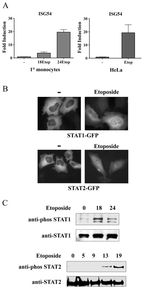
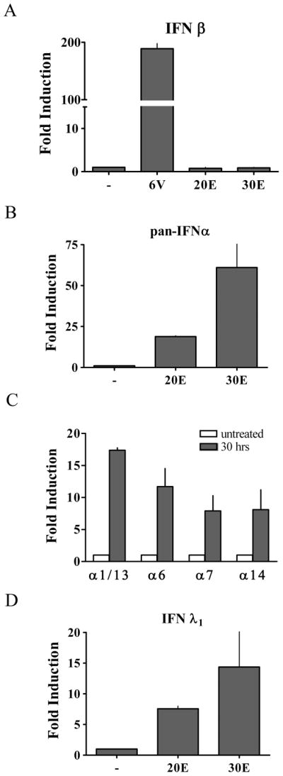
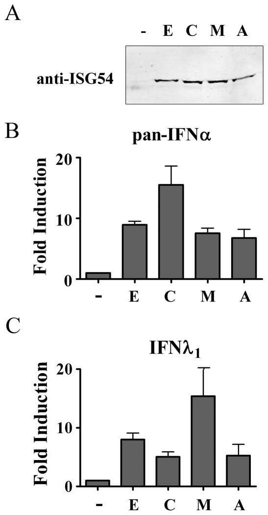
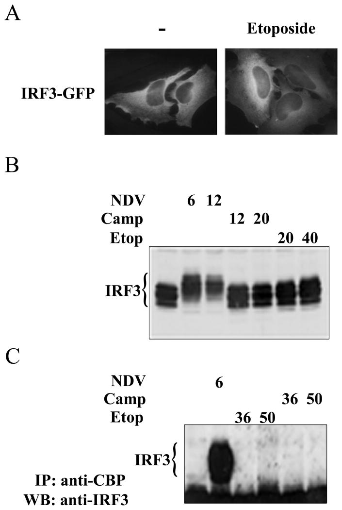
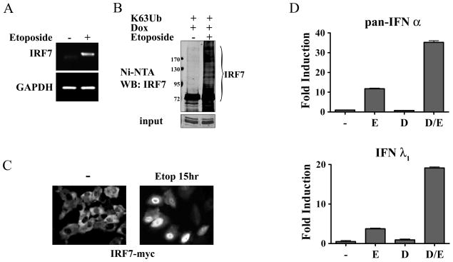
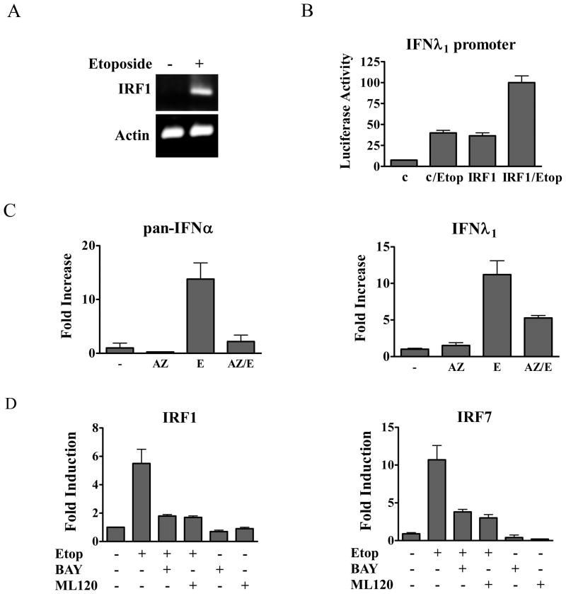
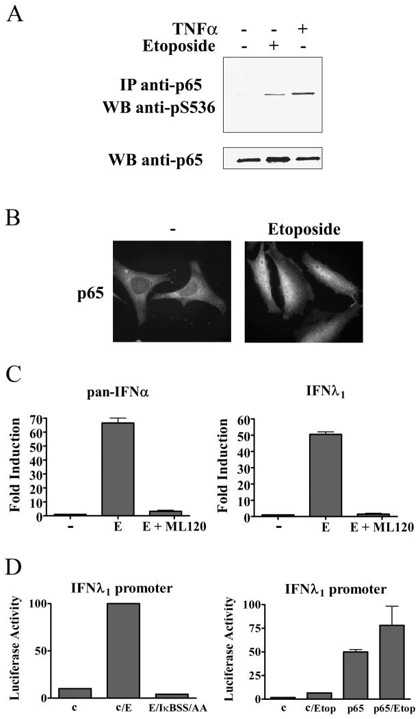
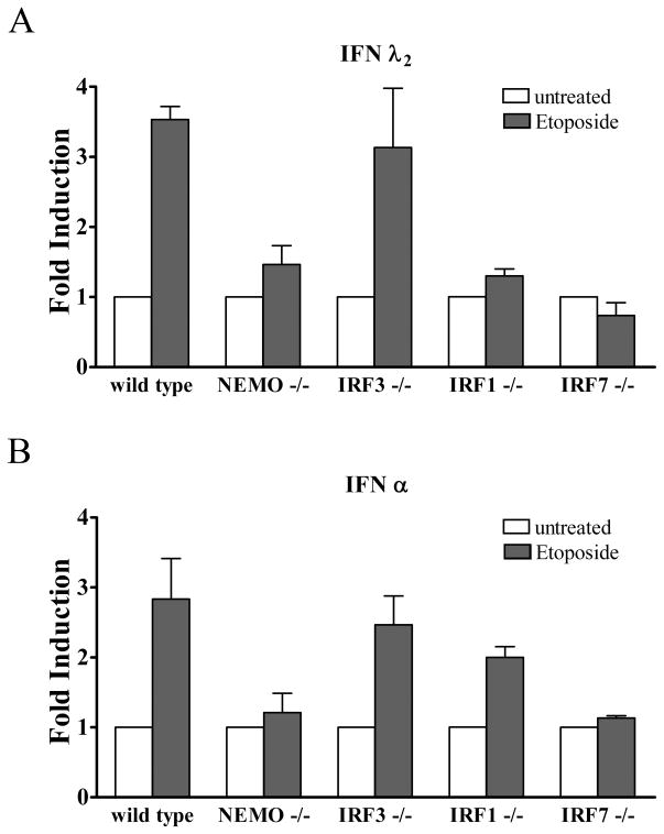
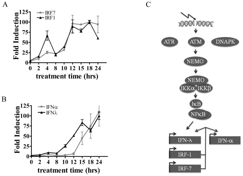
This study reveals a new complexity in the cellular response to DNA damage: activation of IFN signaling. The DNA damage response involves the rapid recruitment of repair enzymes and the activation of signal transducers that regulate cell-cycle checkpoints and cell survival. To understand the link between DNA damage and the innate cellular defense that occurs in response to many viral infections, we evaluated the effects of agents such as etoposide that promote dsDNA breaks. Treatment of human cells with etoposide led to the induction of IFN-stimulated genes and the IFN-α and IFN-λ genes. NF-κB, known to be activated in response to DNA damage, was shown to be a key regulator of this IFN gene induction. Expression of an NF-κB subunit, p65/RelA, was sufficient for induction of the human IFN-λ1 gene. In addition, NF-κB was required for the induction of IFN regulatory factor-1 and -7 that are able to stimulate expression of the IFN-α and IFN-λ genes. Cells that lack the NF-κB essential modulator lack the ability to induce the IFN genes following DNA damage. Breaks in DNA are generated during normal physiological processes of replication, transcription, and recombination, as well as by external genotoxic agents or infectious agents. The significant finding of IFN production as a stress response to DNA damage provides a new perspective on the role of IFN signaling.


Figures

Figure 1
Figure 1. Induction of ISG54 mRNA and activation of STAT signaling in response to etoposide
A) Primary (1°) human monocytes were untreated or treated with etoposide for 0, 18, or 24 hrs. RNA was extracted and real-time RT-PCR was performed with primers specific for ISG54 and quantified (left). HeLa cells were untreated or treated with etoposide for 24 hrs. RNA was extracted and real-time RT-PCR was performed with primers specific for ISG54 and quantified (right). B) HeLa cells were transfected with STAT1-GFP or STAT2-GFP and untreated (−) or treated with etoposide for 24 hours. Fluorescent images are shown. C) HeLa cells were treated with etoposide for the hours indicated and cell lysates were evaluated for endogenous STAT1 (top) or STAT2 (bottom) tyrosine phosphorylation by Western blot. Results represent duplicate determinations in three independent experiments.
Figure 2
Figure 2. Specific IFN species are induced in response to etoposide
A) Primary human monocytes were untreated (−) or treated with etoposide for 20 hours (20E) or 30 hours (30E), or infected with Newcastle Disease Virus (NDV) for 6 hours. mRNA levels were quantified by real-time RT-PCR corresponding to the IFN-β gene in response to etoposide or NDV. B) mRNA levels quantified by pan-specific primers to IFN-α from primary monocytes treated with etoposide as in (A). C) mRNA levels were quantified to specific subspecies IFN-α1/α13, IFN–α6, IFN-α7, and IFN-α14 following 30 hr etoposide treatment of primary monocytes. D) mRNA levels corresponding to the IFN-λ1 gene from primary monocytes treated with etoposide as in (A). Values are means of duplicate determinations in two or three independent experiments.
Figure 3
Figure 3. Various DNA damaging agents activate IFN signaling
THP-1 cells were untreated (−) or treated with 40μg/ml etoposide (E), 5μM camptothecin (C), 20μg/ml mitomycin (M), or 20μg/ml adriamycin (A) for 24 hours. A) Cell lysates were prepared and ISG54 protein levels were evaluated by Western blot. B) IFN-α mRNA induction was evaluated with pan-specific primers and real-time RT-PCR. C) IFN-λ1 specific mRNA expression was evaluated by real-time RT-PCR. Values are means of duplicate determinations in two independent experiments.
Figure 4
Figure 4. IRF-3 is not activated in response to DNA damage
A) HeLa cells were transfected with IRF-3-GFP and left untreated (−) or were treated with etoposide (Etop) for 24 hours. Cellular localization was evaluated by fluorescence microscopy. Images represent random sampling from three independent experiments. B) Cells were untreated or infected with NDV for 6 or 12 hours, treated with camptothecin (Camp) for 12 or 20 hours, or treated with etoposide for 20 or 40 hours. Endogenous IRF-3 was detected in cell lysates by Western blot. C) CBP was immunoprecipitated (IP) from lysates of untreated cells or cells infected with NDV for 6 hours or treated with camptothecin or etoposide for 36 and 50 hours. Western blots (WB) were performed with antibodies to IRF-3.
Figure 5
Figure 5. Evidence for a role of IRF-7 in the IFN response to DNA damage
A) THP-1 cells were untreated (−) or treated with etoposide (+) for 24 hours. IRF-7 mRNA induction was evaluated by RT-PCR and displayed on agarose gels. Faint band in untreated sample is non-specific. mRNA levels of GAPDH are shown as controls. B) HT1080 stable cell line expressing tetracycline inducible IRF-7-Myc-His was transfected with the HA-Ub0R63K ubiquitin (K63Ub), and doxycycline (Dox) was used to induce IRF-7 expression in the absence or presence of etoposide for 24 hours. IRF-7 was collected on nickel charged resins and samples were analyzed by Western blot with antibody to IRF-7. Lower panel shows input before resin with anti-myc antibody. C) Expression of IRF-7-Myc-His was induced with doxycycline in the stable cell line in the absence or presence of etoposide for 15 hours before immunostaining with antibodies to Myc. Imaging analysis of three independent experiments indicate nuclear accumulation of IRF-7 at this time in >50% of the cells. D) Pan-specific primers were used to quantify IFN-α mRNA levels (top) and specific primers were used to assess IFN-λ1 mRNA levels (bottom) in the IRF-7-Myc-His inducible cells by real-time PCR. Cells were untreated (−), treated with etoposide (E), doxycycline (D), or etoposide and doxycycline for 24 hours. Values are means of duplicate determinations in two independent experiments.
Figure 6
Figure 6. Activation and inhibition of IRF and IFN genes
A) THP-1 cells were untreated (−) or treated with etoposide (+) for 24 hours. IRF-1 mRNA levels were evaluated by RT-PCR and displayed on agarose gels. mRNA levels of actin are shown as controls. B) The IFN-λ1 luciferase reporter was expressed in HeLa cells untreated or treated with etoposide (Etop) for 24 hours. Empty vector (c) or IRF-1 expression plasmid (IRF-1) were co-transfected where indicated with or without etoposide treatment and luciferase activity was measured. C) Effects of the ATM inhibitor AZ12622702 (AZ) on IFN-α and IFN-λ gene expression. HeLa cells were untreated or treated with AZ for one hour followed by etoposide (E) for 24 hours as indicated. Real-time PCR was used to quantify IFN-α (left) or IFN-λ (right) mRNA expression. D) Effects of IKKβ inhibitors BAY117085 (BAY) or ML120B (ML120) on IRF-1 and IRF-7 gene expression. HeLa cells were untreated or treated with the inhibitors for one hour followed by etoposide for five hours as indicated. Real-time PCR was used to quantify endogenous IRF-1 (left) or IRF-7 (right) mRNA expression. Quantitative results are means of duplicate determinations in three independent experiments.
Figure 7
Figure 7. NF-κB is activated and required for IFN gene induction in response to etoposide
A) HeLa cells were treated with etoposide for 15 hours or 5ng/ml tumor necrosis factor-α (TNF- α) for 1 hour and p65 was immunoprecipitated (IP) from lysates. Specific antibody to p65 phosphoserine 536 was used for the Western blot (WB). Lower panel displays Western blot with antibody to p65. B) HeLa cells were untreated or treated with etoposide for 2 hours before immunostaining with antibodies to p65. C) HeLa cells were untreated or treated with etoposide (E) in absence or presence of ML120B. left: Real-time PCR was used with pan-specific primers to quantify the endogenous levels of IFN-α mRNA. right: Real-time PCR was used to quantify the endogenous levels of IFN-λ1 mRNA. D) left: The IFN-λ1 luciferase reporter plasmid was co-transfected with empty vector or with a plasmid encoding the dominant negative IκBS32A/S36A (IκBSS/AA) gene. Cells were untreated or treated with etoposide for 24 hours prior to the luciferase assays. right: The IFN-λ1 luciferase reporter plasmid was co-transfected with empty vector (c) or with a plasmid encoding the p65/RelA gene. Cells were untreated or treated with etoposide for 24 hours prior to luciferase assays. Quantitative results are means of duplicate determinations in three independent experiments.
Figure 8
Figure 8
Effect of etoposide on murine embryo fibroblasts. Murine embryo fibroblasts (MEFs) isolated from wt mice, or mice with targeted gene knockouts in NEMO (NEMO−/−), IRF-3 (IRF3−/−), IRF-1 (IRF1−/−), or IRF-7 (IRF7−/−) were untreated or treated with etoposide for 24 hours. Real-time RT-PCR was used to quantify endogenous levels of murine IFNλ2 (top) or pan- IFNα (bottom). Quantitative results are means of duplicate determinations in three independent experiments.
Figure 9
Figure 9. Time course of IRF and IFN mRNA expression in the response to etoposide treatment
THP-1 cells were treated with etoposide, and RNA was isolated from cells during a 24 hour period at the times indicated. Real-time RT-PCR with specific primers was used to quantify IRF-1 and IRF-7 mRNA (A) or IFN-α and IFN-λ1 mRNA (B). Results are means of two experiments performed in duplicate. C) Conceptual model of double strand DNA break response signaling to IFN gene expression. ATM phosphorylation of NEMO promotes its ability to bind and activate the IKK complexes. IKKs phosphorylate IκB releasing NF-κB to activate gene targets IFN-λ1, IRF-1, and IRF-7. The IRFs induce IFN-α genes and enhance IFN-λ1 expression.

References

    1. Vilenchik MM, Knudson AG. Endogenous DNA double-strand breaks: production, fidelity of repair, and induction of cancer. Proc Natl Acad Sci U S A. 2003;100:12871–12876. - PMC - PubMed
    1. Fugmann SD, Lee AI, Shockett PE, Villey IJ, Schatz DG. The RAG proteins and V(D)J recombination: complexes, ends, and transposition. Annu Rev Immunol. 2000;18:495–527. - PubMed
    1. Richardson C, Horikoshi N, Pandita TK. The role of the DNA double-strand break response network in meiosis. DNA Repair (Amst) 2004;3:1149–1164. - PubMed
    1. Sinclair A, Yarranton S, Schelcher C. DNA-damage response pathways triggered by viral replication. Expert Rev Mol Med. 2006;8:1–11. - PMC - PubMed
    1. Mahaney BL, Meek K, Lees-Miller SP. Repair of ionizing radiation-induced DNA double-strand breaks by non-homologous end-joining. Biochem J. 2009;417:639–650. - PMC - PubMed

Publication types

MeSH terms