. 2011 Oct 19;3(105):105ra104.
doi: 10.1126/scitranslmed.3002731.

A small-molecule smoothened agonist prevents glucocorticoid-induced neonatal cerebellar injury

Affiliations

A small-molecule smoothened agonist prevents glucocorticoid-induced neonatal cerebellar injury

Vivi M Heine et al. Sci Transl Med. .

Abstract

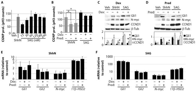
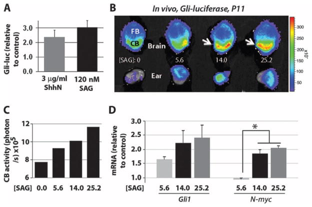
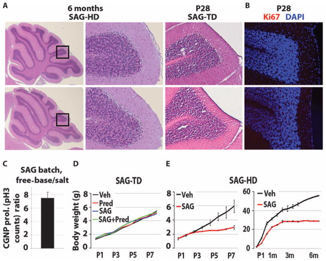
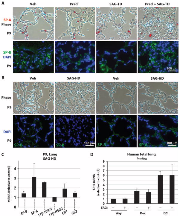
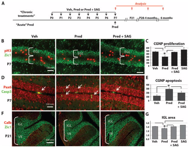
Glucocorticoids are used for treating preterm neonatal infants suffering from life-threatening lung, airway, and cardiovascular conditions. However, several studies have raised concerns about detrimental effects of postnatal glucocorticoid administration on the developing brain leading to cognitive impairment, cerebral palsy, and hypoplasia of the cerebellum, a brain region critical for coordination of movement and higher-order neurological functions. Previously, we showed that glucocorticoids inhibit Sonic hedgehog-Smoothened (Shh-Smo) signaling, the major mitogenic pathway for cerebellar granule neuron precursors. Conversely, activation of Shh-Smo in transgenic mice protects against glucocorticoid-induced neurotoxic effects through induction of the 11β-hydroxysteroid dehydrogenase type 2 (11β-HSD2) pathway. Here, we show that systemic administration of a small-molecule agonist of the Shh-Smo pathway (SAG) prevented the neurotoxic effects of glucocorticoids. SAG did not interfere with the beneficial effects of glucocorticoids on lung maturation, and despite the known associations of the Shh pathway with neoplasia, we found that transient (1-week-long) SAG treatment of neonatal animals was well tolerated and did not promote tumor formation. These findings suggest that a small-molecule agonist of Smo has potential as a neuroprotective agent in neonates at risk for glucocorticoid-induced neonatal cerebellar injury.


Conflict of interest statement

Competing interests: The authors declare that they have no competing interests. The SAG compound was synthesized at Stanford University and is available under a materials transfer agreement.

Figures

Fig. 1
Fig. 1
SAG antagonizes GC effects on cultured primary CGNPs. (A) CGNP proliferation (prol.) stimulated by various SAG concentrations (15 to 240 nM) compared with ShhN (3 μg/ml) and vehicle (Veh) after 24 hours in vitro (n = 4). (B) Effects of dexamethasone (Dex) (40 μM) and prednisolone (Pred) (120 nM) on ShhN-induced (P < 0.001, ANOVA with Tukey’s post hoc; n = 5) and 120 nM SAG–induced CGNP cultures (no significant change). (C and D) Western blots of protein lysates prepared from CGNPs treated with vehicle, ShhN, or 120 nM SAG in the presence or absence of 40 μM dexamethasone (C) [Gli1: P < 0.005, ANOVA; N-myc: P < 0.02, ANOVA (Tukey’s post hoc: SAG versus SAG + dexamethasone, P = 0.02); CCND1: P < 0.005, ANOVA (Tukey’s post hoc: ShhN versus ShhN + dexamethasone, P < 0.0001; SAG versus SAG + dexamethasone, P = 0.05)] or 120 nM prednisolone (D) [Gli1: P < 0.03, ANOVA; N-myc: P < 0.001, ANOVA; CCND1: P < 0.001, ANOVA (Tukey’s post hoc: ShhN versus ShhN + prednisolone, P = 0.04)] for 24 hours (n = 3). Signal intensity of the bands is illustrated in the histograms below. (E and F) Total RNA was isolated from CGNP cultures treated with vehicle, ShhN (E), or 120 nM SAG (F) after 24 hours in the presence or absence of 40 μM dexamethasone or 120 nM prednisolone (n = 3; no significant changes). Asterisks indicate significant changes using Tukey’s post hoc test.
Fig. 2
Fig. 2
SAG activates Gli-luciferase reporter transgene in CGNPs in vitro and in vivo. (A) Gli-luciferase (Gli-luc) reporter expression in primary CGNPs treated with ShhN (3 μg/ml) and SAG (120 nM) for 24 hours in vitro (n = 8). (B) P11 Gli-luciferase mice were injected intraperitoneally with SAG (0, 5.6, 14.0, or 25.2 μg/g) (in saline) and killed after 4 hours. The brains and ears were dissected, placed in luciferin (0.4 mg/ml)/phosphate-buffered saline for 35 min, and then imaged in the Xenogen IVIS detector for 4 min. A photographic image was taken onto which the pseudocolor image representing the spatial distribution of photon count was projected. FB, forebrain; CB, cerebellum. (C) Luciferase levels in the cerebellum quantified in photons per second at various SAG doses. (D) qRT-PCR analysis of SAG effects on the Smo targets Gli1 and N-myc in vivo. SAG dosages (14.0 and 25.2 μg/g) induced significantly increased N-myc levels over a dosage of 5.6 μg/g (P < 0.05, ANOVA with Tukey’s post hoc; n = 2). No significant differences were seen between 14.0 and 25.2 μg/g. Asterisks indicate significant changes with the Tukey’s post hoc test.
Fig. 3
Fig. 3
Results of toxicity studies with SAG at treatment and high doses. (A) Histological analysis of the cerebella of the 6-month-old control and SAG-HD (SAG high dose), and P28 control and SAG-TD (SAG treatment dose)–treated animals. (B) Immunocytochemistry for the proliferation marker Ki67 in the cerebella of P28 control and SAG-TD–treated animals. DAPI, 4′,6-diamidino-2-phenylindole. (C) Determination of treatment and high dose of SAG using CGNP proliferation in vitro. Histogram represents the ratio of the number of pH3-positive cell cultures using free-base SAG versus the salt form of SAG at 1 nM (n = 2). (D and E) Growth curves of vehicle, SAG-TD, prednisolone, prednisolone + SAG-TD (D), and SAG-HD–treated (E) mice.
Fig. 4
Fig. 4
SAG treatment has no detrimental effect on lung maturation. (A and B) Phase-contrast analysis (upper panels) and immunocytochemistry (lower panels) for the differentiation markers SP-B SP-A and nuclear stain DAPI on P9-inflated lungs treated with vehicle, prednisolone, SAG-TD, prednisolone + SAG-TD (A), and SAG-HD (B). (C) qRT-PCR analysis for SP-B, SP-A, 11β-HSD1, 11β-HSD2, Gli1, and Gli2 expression in the P9 lung of SAG-HD–treated mice (n = 3; not significant). Data in (C) are expressed relative to values obtained with vehicle treatment (control), which was set at 1. (D) qRT-PCR for SP-B on human fetal lung explant cultures untreated (Waymouth medium) or treated with dexamethasone (10 nM) or DCI (10 nM dexamethasone, 0.1 mM 8-bromo-cAMP, and 0.1 mM isobutylmethylxanthine together) in the presence or absence of SAG (120 nM) (see Materials and Methods). SAG does not alter the expression level of SP-B (P < 0.005, ANOVA; Tukey’s post hoc: Way-mouth medium versus Waymouth medium + SAG, P > 0.05; dexamethasone versus dexamethasone + SAG, P > 0.05; DCI versus DCI + SAG, P > 0.05; vehicle versus DCI, P < 0.05, n = 3), which is differentially induced by GCs (43). Data are expressed relative to control levels, which were set at 1. β-Actin was used as a reference gene to calculate SP-B expression levels (n = 3). Scale bars, 100 μm [(A) and (B)].
Fig. 5
Fig. 5
SAG protects against neurotoxic effects of prednisolone. (A) Scheme for administration of vehicle, prednisolone, or prednisolone + SAG according to “daily (P0-P7)” or “acute (P7 only)” schedule and histological analysis at P7 or P21. (B to F) Immunocytochemistry of the mitotic marker pH3, apoptosis marker cleaved caspase 3 (Casp3), CGNP markers Zic1 and Pax6, and Purkinje cell marker calbindin (Calb) at P7 and P21. (C) At P7, daily SAG treatment prevented the significant antiproliferative (prednisolone versus prednisolone + SAG: P < 0.02, ANOVA with Tukey’s post hoc, n = 3) and (E) acute SAG treatment proapoptotic (prednisolone versus prednisolone + SAG: P < 0.003, ANOVA with Tukey’s post hoc, n = 3) effects of prednisolone on neonatal CGNPs of the EGL. The prednisolone group is significantly different from all other groups (vehicle, prednisolone + SAG, and SAG). Only the P values of the Tukey’s post hoc test between the prednisolone and the prednisolone + SAG groups are given. (F and G) Significant reduction in the volume of the IGL at P21 induced by prednisolone treatment (P < 0.02, ANOVA with Tukey’s post hoc, n = 3), which was prevented by SAG. Images of lobe VII of the cerebellum shown in (F) are representative of the results in all lobes of the cerebellum. Asterisks indicate significant changes with the Tukey’s post hoc test. Scale bars, 100 μm [(B) and (D)] and 300 μm (F).

Comment in

Similar articles

Cited by

References

    1. Allen MC. Neurodevelopmental outcomes of preterm infants. Curr Opin Neurol. 2008;21:123–128. - PubMed
    1. Doyle LW, Anderson PJ. Adult outcome of extremely preterm infants. Pediatrics. 2010;126:342–351. - PubMed
    1. Yanney M, Marlow N. Paediatric consequences of fetal growth restriction. Semin Fetal Neonatal Med. 2004;9:411–418. - PubMed
    1. Allin MP, Salaria S, Nosarti C, Wyatt J, Rifkin L, Murray RM. Vermis and lateral lobes of the cerebellum in adolescents born very preterm. Neuroreport. 2005;16:1821–1824. - PubMed
    1. Bodensteiner JB, Johnsen SD. Cerebellar injury in the extremely premature infant: Newly recognized but relatively common outcome. J Child Neurol. 2005;20:139–142. - PubMed

Publication types