Comparative Study
. 2011 Nov 15;187(10):5099-113.
doi: 10.4049/jimmunol.1002697. Epub 2011 Oct 19.

Indirect stimulation of human Vγ2Vδ2 T cells through alterations in isoprenoid metabolism

Affiliations
Comparative Study

Indirect stimulation of human Vγ2Vδ2 T cells through alterations in isoprenoid metabolism

Hong Wang et al. J Immunol. .

Abstract

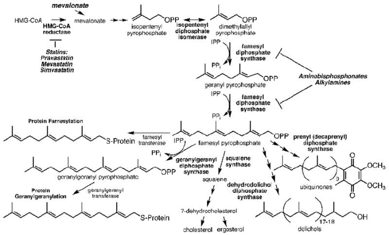
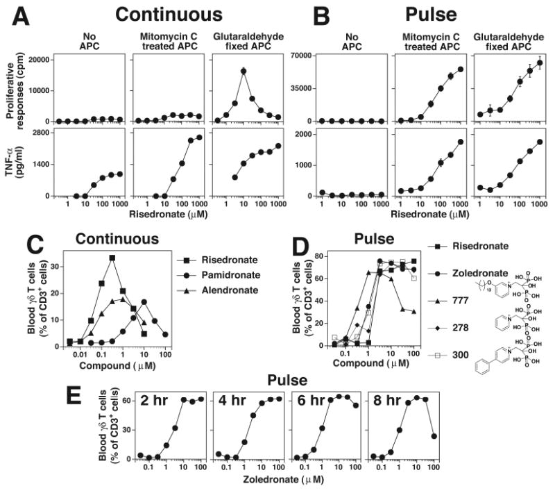
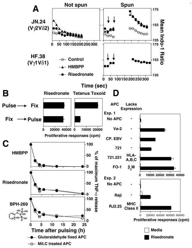
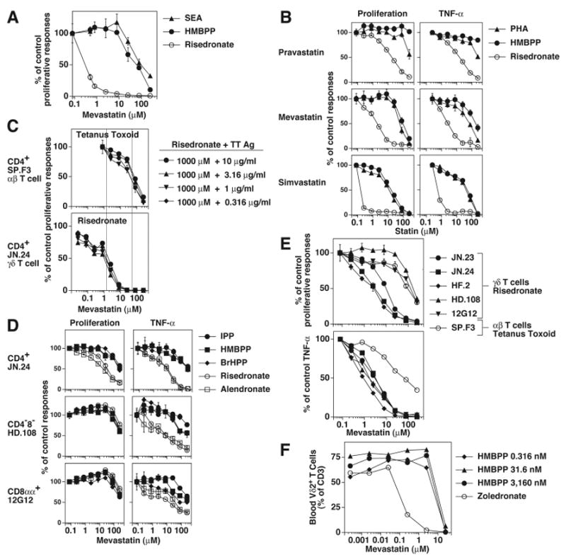
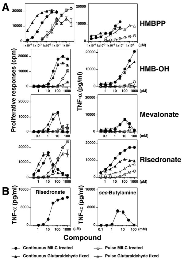
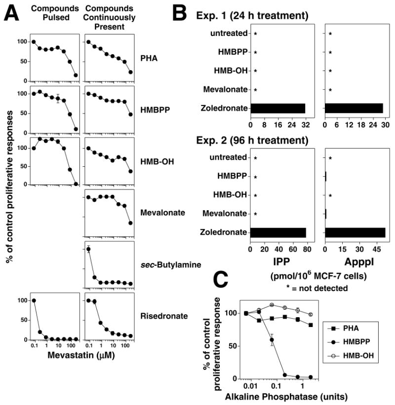
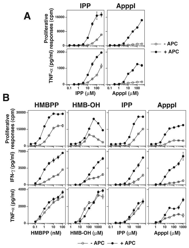
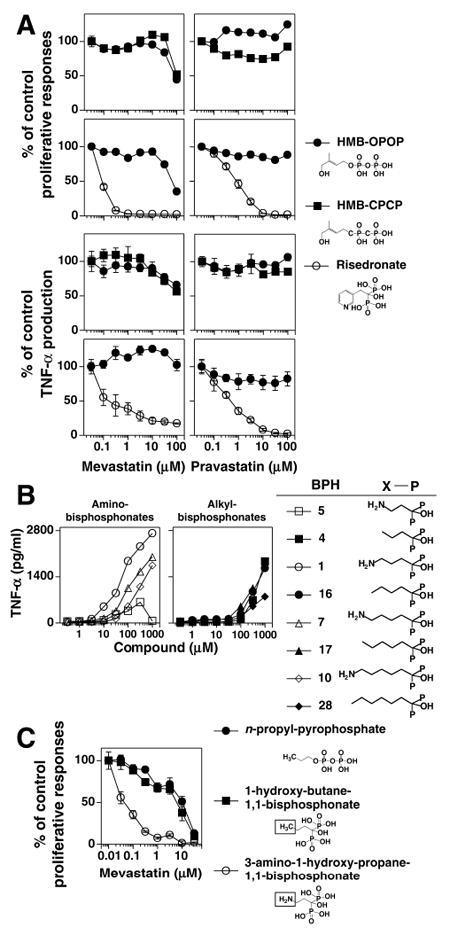
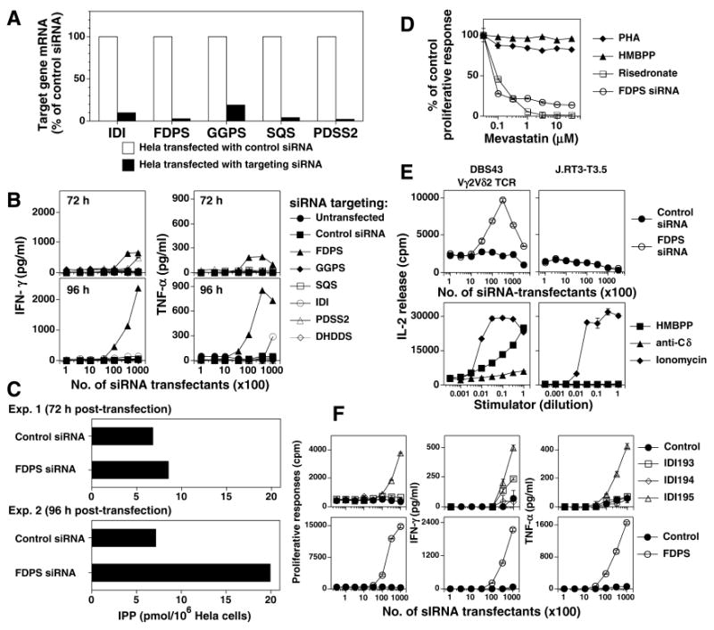
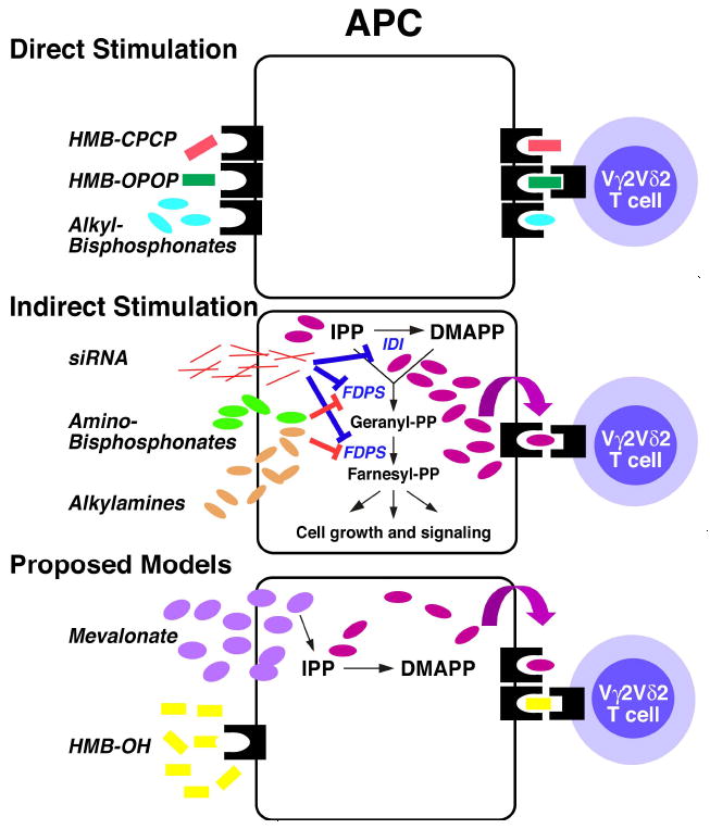
Human Vγ2Vδ2 T cells monitor isoprenoid metabolism by recognizing (E)-4-hydroxy-3-methyl-but-2-enyl pyrophosphate (HMBPP), an intermediate in the 2-C-methyl-d-erythritol-4-phosphate pathway used by microbes, and isopentenyl pyrophosphate (IPP), an intermediate in the mevalonate pathway used by humans. Aminobisphosphonates and alkylamines indirectly stimulate Vγ2Vδ2 cells by inhibiting farnesyl diphosphate synthase (FDPS) in the mevalonate pathway, thereby increasing IPP/triphosphoric acid 1-adenosin-5'-yl ester 3-(3-methylbut-3-enyl) ester that directly stimulate. In this study, we further characterize stimulation by these compounds and define pathways used by new classes of compounds. Consistent with FDPS inhibition, stimulation of Vγ2Vδ2 cells by aminobisphosphonates and alkylamines was much more sensitive to statin inhibition than stimulation by prenyl pyrophosphates; however, the continuous presence of aminobisphosphonates was toxic for T cells and blocked their proliferation. Aminobisphosphonate stimulation was rapid and prolonged, independent of known Ag-presenting molecules, and resistant to fixation. New classes of stimulatory compounds-mevalonate, the alcohol of HMBPP, and alkenyl phosphonates-likely stimulate differently. Mevalonate, a rate-limiting metabolite, appears to enter cells to increase IPP levels, whereas the alcohol of HMBPP and alkenyl phosphonates are directly recognized. The critical chemical feature of bisphosphonates is the amino moiety, because its loss switched aminobisphosphonates to direct Ags. Transfection of APCs with small interfering RNA downregulating FDPS rendered them stimulatory for Vγ2Vδ2 cells and increased cellular IPP. Small interfering RNAs for isopentenyl diphosphate isomerase functioned similarly. Our results show that a variety of manipulations affecting isoprenoid metabolism lead to stimulation of Vγ2Vδ2 T cells and that pulsing aminobisphosphonates would be more effective for the ex vivo expansion of Vγ2Vδ2 T cells for adoptive cancer immunotherapy.


Figures

FIGURE 1
FIGURE 1. Mevalonate pathway and key downstream branches in isoprenoid biosynthesis
HMG-CoA reductase is the rate-controlling enzyme in the mevalonate pathway and is subject to feedback regulation by downstream products. It is also inhibited by statins. FPP synthase converts IPP and DMAPP to GPP and FPP intermediates and is inhibited by aminobisphosphonates and alkylamines. Loss of FPP and GGPP leads to the loss of membrane anchoring of signaling proteins causing signaling defects and, in some cases, apoptosis.
FIGURE 2
FIGURE 2. Aminobisphosphonate toxicity for Vγ2Vδ2 T cell can be avoided by pulsing
A, Continuous culture of Vγ2Vδ2 T cells with aminobisphosphonates inhibits their proliferation but not TNF-α release. Mit. C treated or glutaraldehyde fixed CP.EBV cells were continuously cultured with risedronate and the CD4+ Vγ2Vδ2 T cell clone, JN.23. Supernatants were collected at 16 h for the measurement of TNF-α. The cells were pulsed with 3H-thymidine and harvested 18 h later. B, APC pulsed with risedronate stimulate both proliferation and TNF-α release. Risedronate was pulsed into APC, washed, and then mixed with the CD4+ Vγ2Vδ2 T cell clone, JN.23. TNF-α and cell proliferation were measured as in A. C, Variable expansion of blood Vγ2Vδ2 T cells with continuous exposure to aminobisphosphonates. PBMC were cultured with various aminobisphosphonates for 10 days and Vγ2Vδ2 T cells and CD3+ T cells determined by flow cytometry. D, Consistent blood Vγ2Vδ2 T cell responses to aminobisphosphonates pulsed into monocytes. PBMC were pulsed for 4 hours with the various aminobisphosphonates. The PBMC were then washed and cultured in the presence of IL-2. After 9 days, Vγ2Vδ2 T cells and CD3+ T cells were determined by flow cytometry. E, Expansion of blood Vγ2Vδ2 T cells in PBMC pulsed with zoledronate. PBMC were pulsed for the indicated time with zoledronate, washed, and cultured in the presence of IL-2. After 9 days, Vγ2Vδ2 T cells and CD3+ T cells were determined by flow cytometry.
FIGURE 3
FIGURE 3. Aminobisphosphonate stimulation of Vγ2Vδ2 T cells is a rapid, cell-cell contact-dependent process that is persistent, not inhibited by glutaraldehyde fixation of APC, and does not require known antigen presenting molecules
A, Calcium flux of Vγ2Vδ2 T cells in response to risedronate or HMBPP. The Vγ2Vδ2 T cell clone, JN.24 (top), and the Vγ1Vδ1 T cell clone, HF.38 (bottom), were loaded with indo-1 and calcium flux assessed by flow cytometry. PBS (opened circle), HMBPP (2.5 μM, closed triangle), or risedronate (300 μM, closed circle) was added at the time indicated by the first arrow. Then the cells were either not centrifuged (left) or centrifuged for 50 sec to initiate cell-cell contact (second arrow), incubated for 1 min, and gently resuspended for analysis (right). The mean indo-1 ratio is plotted. B, Glutaraldehyde fixation does not inhibit bisphosphonate stimulation of Vγ2Vδ2 T cells. CP.EBV cells were fixed before or after pulsing with a mix of tetanus toxoid (5 μg/ml) and risedronate (1 mM). The JN.24 Vγ2Vδ2 T cell clone and the SP.F3 αβ T cell clone, that is specific for tetanus toxoid, were then added and cell proliferation measured. C, Time kinetics for the loss of stimulation by pulsed APC. CP.EBV APC were pulsed with HMBPP (3.16 μM), risedronate (316 μM), or the bisphosphonate, BPH-269 (1 mM) followed by the addition of JN.24 T cells at different times. Culture supernatants were harvested 16 hours after T cell addition and TNF-α production assessed by ELISA. Responses at each time point are shown as a percentage of the maximum response. D, Vγ2Vδ2 T cells respond to pulsed risedronate in the absence of classical MHC class I molecules (HLA-A, HLA-B, or HLA-C), MHC class II molecules (HLA-DR, HLA-DQ, HLA-DP, HLA-DMA, or HLA-DMB), CD1 (CD1a, -b, -c, and –d), and β2M-dependent molecules. The JN.24 Vγ2Vδ2 T cell clone was stimulated with APC pulsed with the bisphosphonate, risedronate (1 mM). APC included (Exp. 1) the human fibrosarcoma cell line, Va-2, the EBV B cell lines, CP.EBV and 721 (lacking CD1a, -b, -c, and –d), the mutant EBV line, 721.221 (lacking HLA-A, HLA-B and HLA-C), and the melanoma cell line, FO-1 (lacking β2M) or (Exp. 2) the Burkitt lymphoma, Raji, and its class II-deficient mutant, RJ.2.2.5.
FIGURE 4
FIGURE 4. Indirect stimulation of Vγ2Vδ2 T cells by aminobisphosphonates is more sensitive to statin inhibition than direct stimulation by prenyl pyrophosphates or superantigens
A, Mevastatin inhibition of Vγ2Vδ2 T cell proliferation to staphylococcal enterotoxin A (1 μg/ml), HMBPP (1 μM), or risedronate (10 μM). Mit. C treated CP.EBV cells were pulsed with the above compounds for 1 h, and then cultured with JN.24 T cells. B, Different statins inhibit Vγ2Vδ2 T cell responses. Inhibition by pravastatin, mevastatin, and simvastatin of Vγ2Vδ2 T cell responses to the mitogen, PHA (1:1000), HMBPP (1 μM), or risedronate (1 mM). CP.EBV were preincubated with the indicated statin for 1 h, pulsed with the compounds in the presence of the statin, and then cultured with JN.23 T cells in the presence of the statin. TNF-α and cell proliferation were measured as in Fig. 2A. C, Risedronate-induced Vγ2Vδ2 T cell response is more sensitive to mevastatin inhibition than a tetanus toxoid-induced αβ T cell response presented by the same APC. CP.EBV cells were treated with varying concentrations of mevastatin for 1 h and then pulsed with the mixture of 1 mM risedronate and indicated concentrations of tetanus toxoid. T cells were added to the culture in the presence of mevastatin. After 18 h, the cells were pulsed and harvested 1 day later. D, Mevastatin inhibition of Vγ2Vδ2 T cell responses. The effect of mevastatin on the proliferative and TNF-α responses of the CD4+ γδ T cell clone, JN.24, and the CD4- γδ clones, HD.108 and 12G12 by three prenyl pyrophosphates (100 μM IPP, 1 μM HMBPP, and 10 μM BrHPP) and two bisphosphonates (1mM risedronate and 1 mM alendronate) was determined. E, Mevastatin inhibition of the proliferative and TNF-α responses of five Vγ2Vδ2 T cell clones to risedronate (1 mM) and an αβ T cell clone to tetanus toxoid (10 μg/ml). F, Mevastatin inhibition of blood Vγ2Vδ2 T cell expansion in response to HMBPP or zoledronate. PBMC were incubated with varying concentrations of mevastatin and either HMBPP (3,160-0.316 nM) or zoledronate (3.16 μM) for 6 h, washed, and then cultured with mevastatin. IL-2 was added on day 3. After 8 days, Vγ2Vδ2 T cells and CD3+ T cells were determined by flow cytometry.
FIGURE 5
FIGURE 5. Multiple compounds stimulate Vγ2Vδ2 T cells
A, Stimulation of Vγ2Vδ2 T cells by HMBPP, HMB-OH, mevalonate, and risedronate. The JN.24 Vγ2Vδ2 T cell clone was cultured with non-fixed (mit. C-treated) or fixed Va2 APC that had been pulsed with the indicated compounds or the compounds were added continuously. T cell proliferation and TNF-α release were measured as described in 2A. B, Stimulation of Vγ2Vδ2 T cells by sec-butyamine and risedronate. Untreated CP.EBV and the HF.2 Vγ2Vδ2 T cell clone were cultured continuously with either risedronate or sec-butylamine. Supernatants were collected 16 h later for determination of TNF-α.
FIGURE 6
FIGURE 6. Mevalonate and HMB-OH are relatively resistant to statin inhibition, do not greatly increase intracellular IPP or ApppI levels, and are resistant to alkaline phosphatase
A, Mevastatin inhibition of the response of the CD4+ HF.2 Vγ2Vδ2 T cell clone stimulated (left panels) by APC pulsed with PHA (1:1000), HMBPP (1 μM), HMB-OH (1 mM), or risedronate (1 mM), or (right panels) by APC continuously cultured with PHA (1:1000), HMBPP (1 μM), HMB-OH (1 mM), mevalonate (25 mM), sec-butylamine (5 mM), or risedronate (31.6 μM) continuously present in culture. B, HMB-OH and mevalonate do not greatly increase IPP or ApppI levels. MCF-7 cells were untreated or incubated with HMBPP (10 nM for 24 h or 100 nM for 96 h), HMB-OH (100 μM), or mevalonate (10 mM) for 24 h (Exp. 1, top panels) or 96 h (Exp. 2, bottom panels). For a control, MCF-7 cells were treated for zoledronate (25 μM) for 24 h for both experiments. Cells were then harvested, washed, and lysed with acetonitrile for determination of IPP and ApppI levels by LC/MS (46). C, HMB-OH stimulation is not affected by alkaline phosphatase. Mit. C-treated CP.EBV were cultured in the presence of shrimp alkaline phosphatase continuously with 0.1 μM HMBPP, 1 mM HMB-OH, or 1:4000 diluted PHA or after pulsing with 1 mM risedronate. HF.2 T cells were added and cell proliferation assessed on day 2.
FIGURE 7
FIGURE 7. HMB-OH stimulation of Vγ2Vδ2 T cells in the absence of APC is similar to stimulation by HMBPP and IPP
A, ApppI stimulation is relatively APC dependent. The HF.2 Vγ2Vδ2 T cell clone was cultured with either IPP or ApppI in the presence or absence of mitomycin C-treated CP.EBV. TNF-α and cell proliferation were measured as in Fig. 2A. B, HMB-OH stimulation in the absence of APC is similar to stimulation by HMBPP and IPP. The HF.2 Vγ2Vδ2 T cell clone was cultured with HMBPP, HMB-OH, IPP, or ApppI in the presence or absence of mitomycin C-treated CP.EBV. TNF-α and cell proliferation were measured as in Fig. 2A.
FIGURE 8
FIGURE 8. Linear pyrophosphonates and alkyl-bisphosphonates directly stimulate Vγ2Vδ2 T cells
A, Direct recognition of HMBPP and HMB-CPCP by Vγ2Vδ2 T cells. Mevastatin and pravastatin inhibition of proliferative and TNF-α responses by the HF.2 Vγ2Vδ2 T cell clone stimulated by 0.1 μM of HMB-OPOP or 316 μM of HMB-CPCP continuously present (top panels) or by 1 μM of HMB-OPOP or 1 mM of risedronate that were pulsed with APC (bottom panels). B, Amino- and alkyl-bisphosphonates stimulate Vγ2Vδ2 T cells. Bisphosphonates were tested for their ability to stimulate TNF-α release by the CD4+ JN.23 Vγ2Vδ2 T cell clone. C, Substitution of an amino moiety for carbon 4 in 1-hydroxy-butane-1,1 bisphosphonate switches direct to indirect stimulation. The CD4+ HF.2 Vγ2Vδ2 T cell clone was continuously stimulated by either n-propyl pyrophosphate (20 μM), 1-hydroxy-butane-1,1 bisphosphonate (20 μM), or 3 amino-1-hydroxy-propane-1,1 bisphosphonate (400 μM) in the presence of mevastatin.
FIGURE 9
FIGURE 9. siRNA downregulation of either FDPS mRNA or isopentenyl diphosphate isomerase mRNA in APC results in indirect stimulation of Vγ2Vδ2 T cells with elevations in intracellular IPP levels in APC
A, siRNA treatment greatly decreases mRNA levels of most enzymes in isoprenoid biosynthesis. mRNA levels of enzymes targeted by siRNA were measured in comparison to control siRNA using real-time PCR as detailed in the Materials and Methods. B, Downregulation of FDPS results in APC that stimulate Vγ2Vδ2 T cells. HeLa cells were either untransfected or transfected with control siRNA or siRNA targeting mRNAs for enzymes required for the synthesis of isoprenoid compounds. After 72 h and 96 h, transfected HeLa cells were mixed with HF.2 Vγ2Vδ2 T cells. Supernatants were harvested 16 hours later, and the levels of IFN-γ (left panels) and TNF-α (right panels) determined by ELISA. For each enzyme, 3 siRNAs were tested with the best siRNA shown. Results are representative of 3 experiments. C, Increased intracellular IPP levels in HeLa cells after transfection with siRNA to FDPS. HeLa cells were transfected with either a control siRNA or a siRNA to FDPS. After 72 h or 96 h, the cells were harvested and intracellular IPP level measured. D, Stimulation by APC treated with siRNA to FDPS is sensitive to statin inhibition. HeLa cells were transfected with siRNA to FDPS and after 72 h cultured with HF.2 Vγ2Vδ2 T cells in the presence of mevastatin. For comparison, untransfected HeLa cells were either continuously cultured with 0.1 μM HMBPP or 1:4000 PHA, or pulsed with 1 mM risedronate with HF.2 T cells in the presence of mevastatin. Cultures were pulsed with 1mCi of H-thymidine on day 1 and harvested 16-18 h later. E, Recognition of FDPS siRNA-treated APC is mediated by the Vγ2Vδ2 TCR. The DBS43 Vγ2Vδ2 TCR transfectant or the parent mutant Jurkat cell line, J.RT3-T3.5, was cultured with HeLa cells treated with either a control siRNA or siRNA to FDPS and PMA or with anti-TCRδ1, ionomycin (1 μg/ml), or HMBPP (1 μM) in the presence of Va2 cells and PMA. The supernatants were harvested and IL-2 levels assessed by proliferation of the IL-2-dependent HT-2 cell line. F, Downregulation of isopentenyl diphosphate isomerase (IDI) renders APC stimulatory for Vγ2Vδ2 T cells. Mit. C-treated HeLa cells were transfected with either a control siRNA, 3 different siRNAs targeting IDI, or with a siRNA targeting FDPS. After 96 h, transfected HeLa cells were mixed with HF.2 Vγ2Vδ2 T cells. Culture supernatants were harvested 16 hours later and IFN-γ (middle panels) and TNF-α (right panels) determined by ELISA. Proliferation was assessed on day 2 (left panels).
FIGURE 10
FIGURE 10. Proposed mechanisms for stimulation of Vγ2Vδ2 T cells
Top, Vγ2Vδ2 T cells recognize HMBPP, HMB-CPCP, and alkyl-bisphosphonates presented directly without internalization by an antigen presenting molecule on APC. Middle, aminobisphosphonates and alkylamines indirectly stimulate Vγ2Vδ2 T cells by inhibiting FDPS leading to the accumulation of IPP that can then be presented by the antigen presenting molecule. Transfection of FDPS and IDI siRNA also cause IPP to accumulate and stimulate Vγ2Vδ2 T cells. Bottom, proposed models for mevalonate and HMB-OH. Exogenous mevalonate, a rate limiting intermediate, increases IPP levels modestly that then stimulate Vγ2Vδ2 T cells. In contrast, HMB-OH is likely directly presented as it is relatively APC independent and there is no evidence for IPP accumulation.

Similar articles

Cited by

References

    1. Morita CT, Jin C, Sarikonda G, Wang H. Nonpeptide antigens, presentation mechanisms, and immunological memory of human Vγ2Vδ2 T cells: discriminating friend from foe through the recognition of prenyl pyrophosphate antigens. Immunol Rev. 2007;215:59–76. - PubMed
    1. Shen Y, Zhou D, Qiu L, Lai X, Simon M, Shen L, Kou Z, Wang Q, Jiang L, Estep J, Hunt R, Clagett M, Sehgal PK, Li Y, Zeng X, Morita CT, Brenner MB, Letvin NL, Chen ZW. Adaptive immune response of Vγ2Vδ2+ T cells during mycobacterial infections. Science. 2002;295:2255–2258. - PMC - PubMed
    1. Wang L, Kamath A, Das H, Li L, Bukowski JF. Antibacterial effect of human Vγ2Vδ2 T cells in vivo. J Clin Invest. 2001;108:1349–1357. - PMC - PubMed
    1. Wilhelm M, Kunzmann V, Eckstein S, Reimer P, Weissinger F, Ruediger T, Tony H-P. γδ T cells for immune therapy of patients with lymphoid malignancies. Blood. 2003;102:200–206. - PubMed
    1. Dieli F, Vermijlen D, Fulfaro F, Caccamo N, Meraviglia S, Cicero G, Roberts A, Buccheri S, D’Asaro M, Gebbia N, Salerno A, Eberl M, Hayday AC. Targeting human γδ T cells with zoledronate and interleukin-2 for immunotherapy of hormone-refractory prostate cancer. Cancer Res. 2007;67:7450–7457. - PMC - PubMed

Publication types

MeSH terms