. 2012 Feb;40(4):1717-27.
doi: 10.1093/nar/gkr855. Epub 2011 Oct 19.

RecA homology search is promoted by mechanical stress along the scanned duplex DNA

Affiliations

RecA homology search is promoted by mechanical stress along the scanned duplex DNA

Claudia Danilowicz et al. Nucleic Acids Res. 2012 Feb.

Abstract

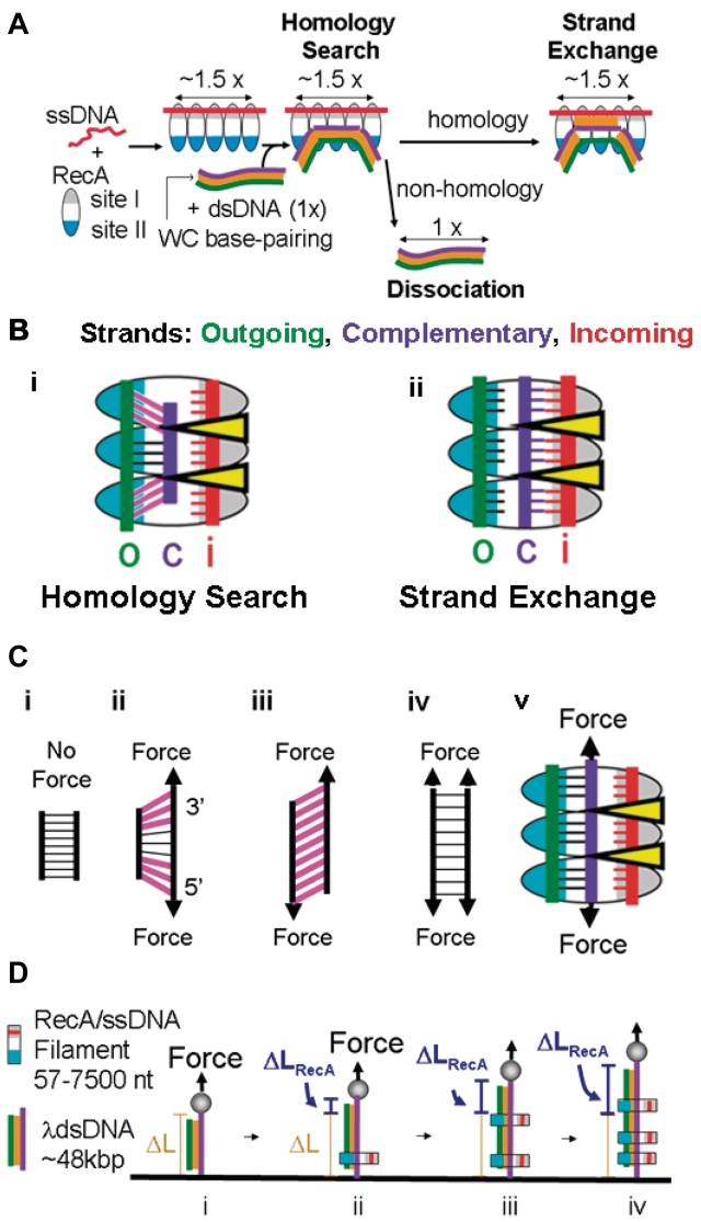
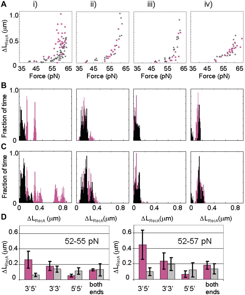
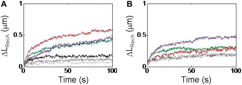
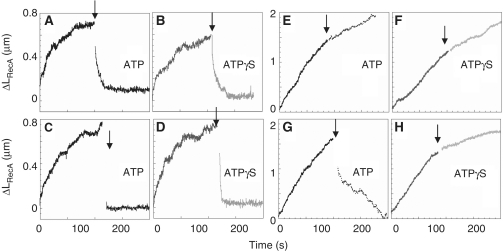
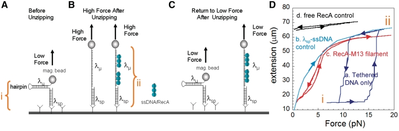
A RecA-single-stranded DNA (RecA-ssDNA) filament searches a genome for sequence homology by rapidly binding and unbinding double-stranded DNA (dsDNA) until homology is found. We demonstrate that pulling on the opposite termini (3' and 5') of one of the two DNA strands in a dsDNA molecule stabilizes the normally unstable binding of that dsDNA to non-homologous RecA-ssDNA filaments, whereas pulling on the two 3', the two 5', or all four termini does not. We propose that the 'outgoing' strand in the dsDNA is extended by strong DNA-protein contacts, whereas the 'complementary' strand is extended by the tension on the base pairs that connect the 'complementary' strand to the 'outgoing' strand. The stress resulting from different levels of tension on its constitutive strands causes rapid dsDNA unbinding unless sufficient homology is present.


Figures

Figure 1.
Figure 1.
RecA–ssDNA binding to dsDNA. (A) Schematic representation of RecA-mediated reactions: incoming ssDNA (red line), outgoing strand (green line), complementary strand (purple line), Watson–Crick pairing (orange) and RecA: site I (gray region of oval) and site II (blue region of oval). (B) Schematic representation of the extension and tension on dsDNA bound to RecA–ssDNA filaments with pink highlighting base pairs under tension and yellow triangles indicating regions occupied by the L1 and L2 loops and their attached alpha helices which interact strongly with the incoming strand (i) dsDNA bound to the pre-synaptic filament (ii) dsDNA bound to a RecA–ssDNA filament in the post-strand exchange state (C) Schematic representation of the extension and tension on dsDNA with the same color scheme used in (B) (i) in the absence of external force (ii) with external force applied to the 3′5′-ends of one strand (iii) shear force applied to opposite ends of opposite strands (3′3′ or 5′5′ pulling) (iv) with external force applied to both strands at both ends (v) with external force applied to the 3′5′-ends of the complementary strand in the homology searching complex (D) Schematic representation of the extension-based assay for measuring the extension of dsDNA due to the binding of RecA–ssDNA filaments (i) λ dsDNA tethered between a capillary tube and a magnetic bead under force (arrow) that is extended without [ΔL, (i)] and with [ΔLRecA (ii–vi)] binding of non-homologous RecA–ssDNA filaments.
Figure 2.
Figure 2.
RecA–ssDNA filaments bind to non-homologous dsDNA pulled by 3′5′ ends. Confocal microscope visualization (linearly enhanced for brightness and contrast) of fluorescent RecA–ssDNA filaments (white arrows) bound to λ dsDNA. The force applied to the bead is indicated by the yellow arrows.
Figure 3.
Figure 3.
RecA–ssDNA filaments extend non-homologous dsDNA pulled by 3′5′ ends. (A) ΔLRecA's for λ dsDNAs controls with free ssDNA or λ dsDNA only (red, N = 87) and λ dsDNAs with RecA–ssDNA filaments (blue, N = 454). (B) Single molecule extension profiles for non-homologous binding. Different colors are used to represent the response for each different single molecule. The curves ending at positions corresponding to 1, 3 and 4 filaments are 57.0, 59.0 and 60.9 pN, respectively. The red and green curves ending at two-filament lengths correspond to 58.9 and 57.2 pN, respectively. The filaments were prepared in a buffer containing ATPγS, but measured in a buffer containing ATP Pauses at full filament lengths are indicated by horizontal bars the same color as the curve. (C) ΔLRecA probability distributions for periods of ≥2 s constant λ dsDNA length, for all filaments of 400–800 nt at 50–62 pN. (D) DNA extensions within homology search complexes on dsDNA (color) or λ ssDNA.
Figure 4.
Figure 4.
Binding of RecA–ssDNA filaments to dsDNA pulled from different ends. (A) ΔLRecA versus force for 1000-nt filaments (magenta circles). λ dsDNAs are pulled on (i) 3′5′-ends of one strand, (ii) 3′3′-ends, (iii) 5′5′-ends, (iv) both ends of each of the two constituent strands of the dsDNA, respectively, and controls for each pulling technique in the absence of RecA (gray triangles). (B) Histograms of the fraction of the total number of beads that showed a particular ΔLRecA after a force has been applied for 60 s (magenta) for dsDNA pulled (from left to right) from 3′5′-ends of one strand, 3′3′ends, 5′5′-ends, and both ends of each of the two constituent strands of the dsDNA; force range: 52-55 pN and negative controls shown in gray. (C) Histograms of the fraction of the total number of beads that showed a particular ΔLRecA after a force has been applied for 60 s (magenta) for dsDNA pulled (from left to right) from 3′5′-ends of one strand, 3′3′-ends, 5′5′-ends and both ends of each of the two constituent strands of the dsDNA: force range: 52–57 pN; negative controls shown in gray. (D) Bar graphs for ΔLRecA values between 52 and 55 pN and 52 and 57 pN, for 1000-nt filaments. λ dsDNAs are pulled 3′5′-, 3′3′-, 5′5′- and both ends of both strands: positives (magenta) and controls (gray). In these experiments filaments were prepared in a buffer containing ATPγS and measured in a buffer containing ATPγS.
Figure 5.
Figure 5.
Single molecule extension profiles for non-homologous RecA–ssDNA (1 kb) filaments binding when force is applied to the 3′5′-ends of one of the constituent strands in RecA buffer pH 7.6 containing ATPγS. (A) Force applied to the same constituent strand as in Figure 3. Green curve: 55.7 pN, red: 59.1 pN, and purple: 58.4 pN. Control curves are black (56.6 pN), dark gray (59.8 pN) and light gray (57.5 pN). (B) Force was applied to the 3′5′-ends of the other constituent strand. Green: 56 pN, red: 59.6 pN, and purple: 58.4 pN. Control curves are light gray (59 pN) and dark gray (57.5 pN).
Figure 6.
Figure 6.
Non-homologous binding is stabilized by external force. Preformed RecA–ssDNA filaments (A–D) or free RecA (E–H) added to λ dsDNAs. After 100 s at 57 pN, force was reduced (arrow) by 10 to 47 pN (A, B, E, F) or 40 to 17 pN (C, D, G, H). Non-homologous RecA–ssDNA binding is rapidly lost if force is reduced, irrespective of ATP hydrolysis (A, D). RecA binding (via site I) continues in ATPγS (F, H) and is lost in ATP if force is reduced <30 pN (E, G).
Figure 7.
Figure 7.
Interaction of RecA–ssDNA filaments with ssDNA during DNA ‘unzipping’. Representation of unzipping experiment: One λ dsDNA molecule (λsp) acts as spacer and the second λ dsDNA (λµ) is unzipped by force. λµ contains a hairpin connecting its two constituent ssDNA strands at one terminus and a magnetic bead at the free terminus between λsp and λµ. i is the extension distance for forces between ∼ 2 and 12 pN at which λsp B-form duplex molecule is stretched out, but the λµ molecule remains fully zipped. ii is the extension distance at which λµ is fully unzipped, but the λsp remains in B-form. (A) The initial extension before unzipping. (B) The extension after unzipping while a force >15 pN is maintained. (C) The extension after the force has been lowered to ∼5 pN. (D) Extension versus force curves for unzipping. The force was first increased and then decreased. (a) No added protein or nucleoprotein filaments (b) ‘unrezippable’ control curve constructed by taking the sum of the extension versus force curves for λsp alone and two times that for a λ ssDNA strand obtained by thermal melting of dsDNA. (Note: in this case rezipping is impossible because the complementary strand is not present.) (c) Unzipping in the presence of RecA–ssDNA filaments (M13 ssDNA). (d) Unzipping in the presence of free RecA protein. In (a), (c) and (d) curves generated by increasing force differed from those generated by decreasing force. In these cases, upturned arrowheads mark the ascending curves and down-turned arrowheads the descending ones.

Similar articles

Cited by

References

    1. Kowalczykowski SC, Eggleston AK. Homologous pairing and DNA strand-exchange. Annu. Rev. Biochem. 1994;63:991–1043. - PubMed
    1. Roca AI, Cox MM. The RecA protein: structure and function. Crit. Rev. Biochem. Mol. Biol. 1990;25:415–456. - PubMed
    1. Howard-Flanders P, West SC, Stasiak A. Role of RecA protein spiral filaments in genetic recombination. Nature. 1984;309:17–23. - PubMed
    1. Müller B, Koller T, Stasiak A. Characterization of the DNA binding activity of stable RecA-DNA complexes. Interaction between the two DNA binding sites within RecA helical filaments. J. Mol. Biol. 1990;212:97–112. - PubMed
    1. Takahashi M, Kubista M, Nordén B. Binding stoichiometry and structure of RecA-DNA complexes studied by flow linear dichroism and fluorescence spectroscopy. Evidence for multiple heterogeneous DNA co-ordination. J. Mol. Biol. 1989;205:137–147. - PubMed

Publication types