

Evaluation of brain tumor vessels specific contrast agents for glioblastoma imaging
- PMID: 22013169
- PMCID: PMC3245996
- DOI: 10.1093/neuonc/nor183
Evaluation of brain tumor vessels specific contrast agents for glioblastoma imaging
Abstract
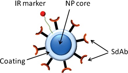
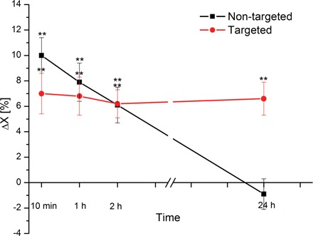
A mouse model of glioblastoma multiforme was used to determine the accumulation of a targeted contrast agent in tumor vessels. The contrast agent, consisting of superparamagnetic iron oxide coated with dextran, was functionalized with an anti-insulin-like-growth-factor binding protein 7 (anti-IGFBP7) single domain antibody. The near infrared marker, Cy5.5, was also attached for an in vivo fluorescence study. A 9.4T magnetic resonance imaging (MRI) system was used for in vivo studies on days 10 and 11 following tumor inoculation. T(2) relaxation time was used to measure the accumulation of the contrast agent in the tumor. Changes in tumor to brain contrast because of active targeting were compared with a nontargeted contrast agent. Effective targeting was confirmed with near infrared measurements and fluorescent microscopic analysis. The results showed that there was a statistically significant (P < .01) difference in normalized T(2) between healthy brain and tumor tissue 10 min, 1 h, and 2 h point postinjection of the anti-IGFBP7 single domain antibody targeted and nontargeted iron oxide nanoparticles. A statistical difference remained in animals treated with targeted nanoparticles 24 h postinjection only. The MRI, near infrared imaging, and fluorescent microscopy studies showed corresponding spatial and temporal changes. We concluded that the developed anti-IGFBP7-iron oxide single domain antibody-targeted MRI contrast agent selectively binds to abnormal vessels within a glioblastoma. T(2)-weighted MRI and near infrared imaging are able to detect the targeting effects in brain tumors.
Figures


 MRI of a CD-1 nude brain tumor mouse model. (A) Before, (B) 20 min, (C) 1 h, and (D) 24 h postinjection of targeted (top row) and nontargeted (bottom row) superparamagnetic Fe3O4 nanoparticles. A gradient echo flow compensation method was used with the following parameters: FOV = 2 × 2 cm, slice thickness 1 mm, TR = 50 ms, TE = 7 ms, 50 kHz bandwidth, 15 degree flip angle and matrix size 128 × 128.


References
-
- Sutherland GR, Florell R, Choi N, Sima AA. Epidemiology of primary intracranial neoplasms in Manitoba, Canada. Can J Neurol Sci. 1987;14:586–592. - PubMed
-
- Ohagaki H, Kleihues P. Population based studies on incidence, survival rates, and genetic alterations in astrocytic and oligodendrglial gliomas. J Neuropathol Exp Neurol. 2005;64(6):479–489. - PubMed
Publication types
MeSH terms
Substances
Grants and funding
LinkOut - more resources
Full Text Sources
Other Literature Sources
Medical
Miscellaneous
