. 2011 Dec 15;204(12):1902-11.
doi: 10.1093/infdis/jir663. Epub 2011 Oct 19.

Evaluation of monkeypox disease progression by molecular imaging

Affiliations

Evaluation of monkeypox disease progression by molecular imaging

Julie Dyall et al. J Infect Dis. .

Abstract

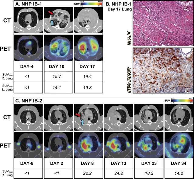
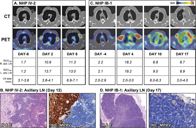
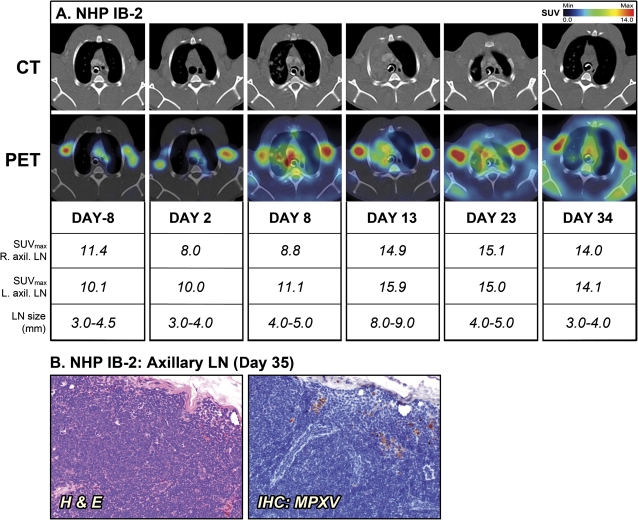
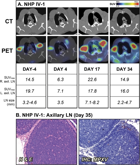
Infection of nonhuman primates (NHPs) with monkeypox virus (MPXV) is currently being developed as an animal model of variola infection in humans. We used positron emission tomography and computed tomography (PET/CT) to identify inflammatory patterns as predictors for the outcome of MPXV disease in NHPs. Two NHPs were sublethally inoculated by the intravenous (IV) or intrabronchial (IB) routes and imaged sequentially using fluorine-18 fluorodeoxyglucose ((18)FDG) uptake as a nonspecific marker of inflammation/immune activation. Inflammation was observed in the lungs of IB-infected NHPs, and bilobular involvement was associated with morbidity. Lymphadenopathy and immune activation in the axillary lymph nodes were evident in IV- and IB-infected NHPs. Interestingly, the surviving NHPs had significant (18)FDG uptake in the axillary lymph nodes at the time of MPXV challenge with no clinical signs of illness, suggesting an association between preexisting immune activation and survival. Molecular imaging identified patterns of inflammation/immune activation that may allow risk assessment of monkeypox disease.


Figures

Figure 1.
Figure 1.
PET/CT) imaging of lung inflammation. A, For the nonhuman primate (NHP) IB-1 that succumbed to infection, sequential axial lung sections were imaged on day –4 and days 10 and 17 after infection. B, Histopathology and monkeypox virus (MPXV) antigen reactivity of lung on day 17. C, Sequential axial lung sections were imaged in surviving NHP IB-2 on day –8 and days 2, 8, 13, 23, and 34 after infection. (Red arrow) Hazy attenuation. (Blue arrow) Consolidated area. Maximum standard uptake values (SUVmax) for fluorine-18 fluorodeoxyglucose (18F-FDG) in right and left sections of lung were determined on scanning days by PET. The PET/CT slice that was chosen for each image corresponds to the maximum SUV value determined in the volume of interest (VOI) of the lung. Abbreviations: H & E, hematoxylin and eosin; IB, intrabronchial; IHC, immunohistochemistry.
Figure 2.
Figure 2.
Sequential positron emission tomography/computed tomography (PET/CT) imaging of axillary lymph nodes (LNs) in nonhuman primates (NHPs) that succumbed to disease. A, NHP IV-2 was scanned on day –8 and days 2 and 8 after infection (d12, day of death). B, Histopathology of NHP IV-2 on day 12. C, NHP IB-1 was scanned on days –4 and days 10 and 17 after infection (day 17, day of death). D, Histopathology of NHP IB-1 on day 17. Fluorine-18 fluorodeoxyglucose (18F-FDG) maximum standard uptake values (SUVmax) and short axis measurements of axillary LNs (LN size) were determined on scan days by PET and CT imaging, respectively. The PET/CT slice that was chosen corresponds to the maximum SUV value of the LNs. Abbreviations: H & E, hematoxylin and eosin; IB, intrabronchial; IHC, immunohistochemistry; IV, intravenous; MPXV, monkeypox virus.
Figure 3.
Figure 3.
Positron emission tomography/computed tomography (PET/CT) imaging of axillary lymph nodes (LNs) in the surviving nonhuman primate (NHP) IB-2. A, Fluorine-18 fluorodeoxyglucose (18FDG)–PET/CT scans were performed on days –8, 2, 8, 13, 23, and 34 of the study. Maximum standard uptake values (SUVmax) for 18F-FDG uptake and short axis measurements of axillary LNs (LN size) were determined on scan days by PET and CT imaging, respectively. B, Histopathology and monkeypox virus (MPXV) antigen reactivity of axillary LN on day 35. The PET/CT slice that was chosen for each image corresponds to the maximum SUV value determined in the volume of interest (VOI) of the LNs. Abbreviations: H & E, hematoxylin and eosin; IB, intrabronchial; IHC, immunohistochemistry.
Figure 4.
Figure 4.
Positron emission tomography/computed tomography (PET/CT) imaging of axillary lymph nodes (LNs) in the surviving nonhuman primate (NHP) IV-1. A, Fluorine-18 fluorodeoxyglucose (18FDG)–PET/CT scans were performed on days –4, 4, 17, and 34 of the study. Maximum standard uptake values (SUVmax) for 18F-FDG uptake and short axis measurements of axillary LNs (LN size) were determined on scan days by PET and CT imaging, respectively. B, Histopathology and monkeypox virus (MPXV) antigen reactivity of axillary LN on day 35. The PET/CT slice that was chosen for each image corresponds to the maximum SUV value determined in the volume of interest (VOI) of the LNs. Abbreviations: H & E, hematoxylin and eosin; IHC, immunohistochemistry; IV, intravenous.

Similar articles

Cited by

References

    1. Likos AM, Sammons SA, Olson VA, et al. A tale of two clades: monkeypox viruses. J Gen Virol. 2005;86:2661–72. - PubMed
    1. Heymann DL, Szczeniowski M, Esteves K. Re-emergence of monkeypox in Africa: a review of the past six years. Br Med Bull. 1998;54:693–702. - PubMed
    1. Learned LA, Reynolds MG, Wassa DW, et al. Extended interhuman transmission of monkeypox in a hospital community in the Republic of the Congo, 2003. Am J Trop Med Hyg. 2005;73:428–34. - PubMed
    1. Parker S, Nuara A, Buller RM, Schultz DA. Human monkeypox: an emerging zoonotic disease. Future Microbiol. 2007;2:17–34. - PubMed
    1. Huhn GD, Bauer AM, Yorita K, et al. Clinical characteristics of human monkeypox, and risk factors for severe disease. Clin Infect Dis. 2005;41:1742–51. - PubMed

Publication types

MeSH terms

Substances