. 2011 Jul;21(3):170-5.
doi: 10.4103/0971-3026.85363.

Recent advances in intestinal imaging

Affiliations

Recent advances in intestinal imaging

Rakesh Sinha. Indian J Radiol Imaging. 2011 Jul.

Abstract

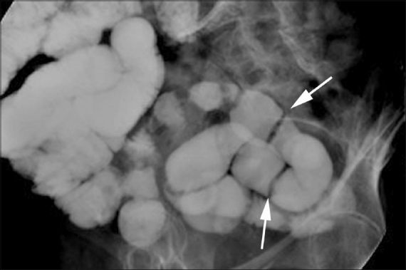
In recent years, advances in scanner technology and competition from other specialties have produced rapid changes in the way the intestines are imaged. MRI and CT scan along with the traditional enteroclysis examination have emerged at the forefront of intestinal imaging. Functional modalities such as diffusion and perfusion imaging are also changing the way tumors and inflammatory bowel diseases are evaluated. CT colonography is now a valid alterative to optical colonoscopy. Contrast-enhanced USG is being used for the assessment of inflammation and post-treatment changes. In this review, recent advances in intestinal imaging are described.

Keywords: Computed tomography; intestines; magnetic resonance imaging.


Conflict of interest statement

Conflict of Interest: None declared.

Figures

Figure 1
Figure 1
Barium–methylcellulose bowel examination shows linear filling defects (webs) (arrows) in the distal ileum secondary to the use of nonsteroidal anti-inflammatory drugs (NSAIDs)
Figure 2
Figure 2
Magnified view from an air–barium double contrast enteroclysis examination shows early linear mucosal breaks and ulcers (arrow) in a patient with proven Crohn disease
Figure 3
Figure 3
Axial CT enteroclysis examination demonstrates a segment of kinked bowel (arrowhead) and several adhesive bands across other segments (arrows). This patient had undergone several negative CT examinations previously
Figure 4 (A,B)
Figure 4 (A,B)
Screenshot (A) from a CT colonographic examination shows a polyp in the colon. Top two axial images show the location of the polyp within a marker box, whereas the bottom two are virtual 3D images in the supine and prone positions which detail polyp size and distance from the rectum. Mucosal views (B) show a virtual dissection view of the colon with a small polyp (arrow) in the left image. On the right, translucency rendering shows this lesion to be homogenously dense (red), implying this is retained fecal matter
Figure 5
Figure 5
A patient with Crohn disease. MRI enterography examination shows good opacification of the small and large bowel with thickening of the inflamed cecal wall (arrow)
Figure 6
Figure 6
High-resolution USG image shows an inflamed bowel segment, with marked enhancement of the mucosal vessels (arrowheads) and engorgement of the penetrating blood vessels (arrow)
Figure 7
Figure 7
PET/CT image shows a recurrent rectal tumor as a hot spot (arrow) just anterior to the presacral fascia
Figure 8
Figure 8
A patient with rectal cancer. Diffusion-weighted MRI image shows high signal in the cancer tissue (arrow) and involved lymph node in the mesorectum (short arrow)

Similar articles

Cited by

References

    1. Balthazar EJ, Herlinger H, Maglinte D, Birnbaum BA. 2nd ed. Germany: Springer; 2001. Clinical Imaging of the Small Intestine.
    1. Maglinte DD, Kelvin FM, O’Connor K, Lappas JC, Chernish SM. Current status of small bowel radiography. Abdom Imaging. 1996;21:247–57. - PubMed
    1. Maglinte DDT. Small bowel imaging: A rapidly changing field and a challenge to radiology. Eur Radiol. 2006;16:967–71. - PubMed
    1. Maglinte D, Sandrasegaran K, Tann M. Advances in alimentary tract imaging. World J Gastroenterol. 2006;12:3139–45. - PMC - PubMed
    1. Maglinte D, Sandrasegaran K, Chiorean M, Dewitt J, McHenry L, Lappas J. Radiologic investigations complement and add diagnostic information to capsule endoscopy of small-bowel diseases. AJR Am J Roentgenol. 2007;189:306–12. - PubMed