

doi: 10.4103/0971-3026.85364.
CT features, mimics and atypical presentations of gastrointestinal stromal tumor (GIST)
Affiliations
- PMID: 22013291
- PMCID: PMC3190488
- DOI: 10.4103/0971-3026.85364
Item in Clipboard
CT features, mimics and atypical presentations of gastrointestinal stromal tumor (GIST)
Indian J Radiol Imaging.
2011 Jul.
Abstract
The term stromal tumor was coined in 1983 by Clark and Mazur for smooth muscle neoplasm of the gastrointestinal tract (GIT). Gastrointestinal stromal tumors (GIST) are nonepithelial tumors arising from the interstitial cells of Cajal, which express KIT protein-CD117 on immunohistochemistry. GIST can arise anywhere in the GIT, including the mesentery, omentum, and retroperitoneum.
Keywords: Exophytic; GIST; immunohistochemistry.
Conflict of interest statement
Figures
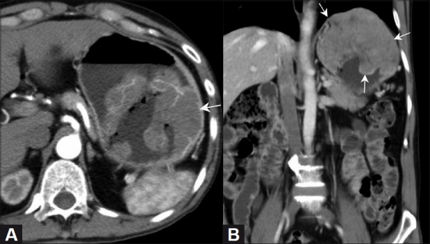
Gastric GIST. Axial (A) and coronal (B) contrast-enhanced CT scan in a 70-year-old male show a well-defined heterogeneously enhancing mass (arrow) in the stomach, with intraluminal and exophytic components, better defined on the coronal images. The left hemidiaphragm is raised
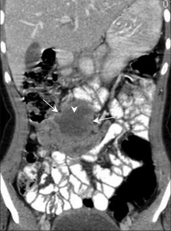
Jejunal GIST. Coronal contrast-enhanced CT scan in a 16-year-old girl shows an exophytic bowel mass (arrow) arising from the jejunal loops. There are areas of necrosis (arrowhead) but there is no evidence of bowel obstruction
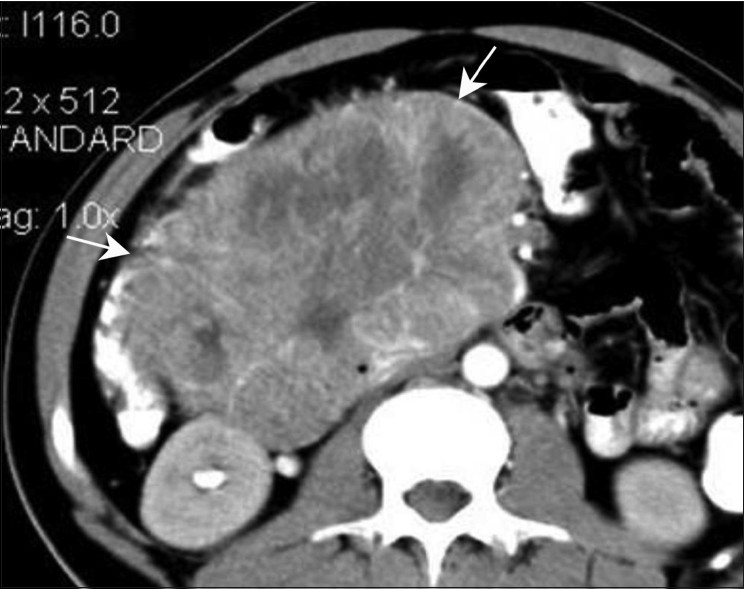
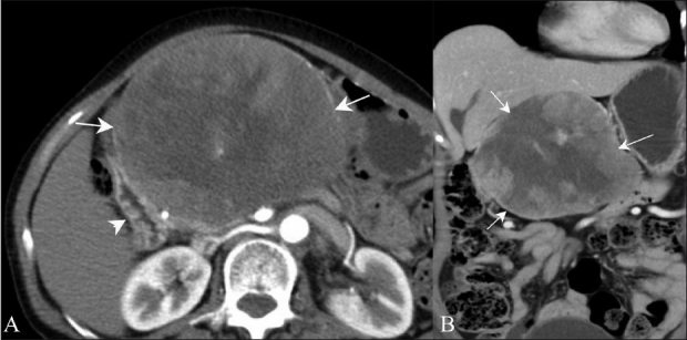
Duodenal GIST. Axial contrast-enhanced CT scan in a 35-year-old female show a well-defined heterogeneously enhancing mass (arrow) along the second part of the duodenum
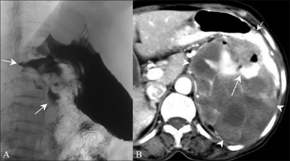
Malignant GIST. Barium meal study (A) shows an irregular filling defect along the fundus and greater curvature of the stomach, suggestive of ulceration (arrow). Axial contrast-enhanced CT scan (B) shows an exophytic heterogeneously enhancing mass (arrowhead) arising from the stomach (small arrow) with areas of ulceration (long arrow)
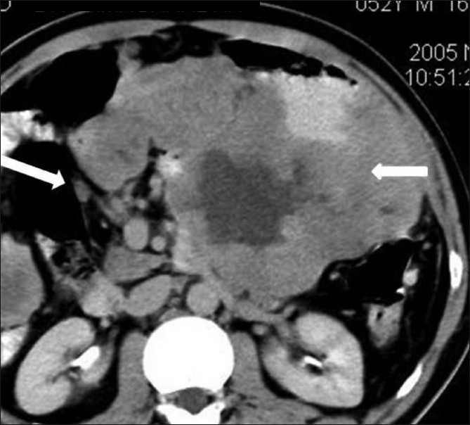
Peritoneal GIST. Axial contrast-enhanced CT scan in 65-year-old male shows multiple heterogeneously enhancing peritoneal masses (arrows)
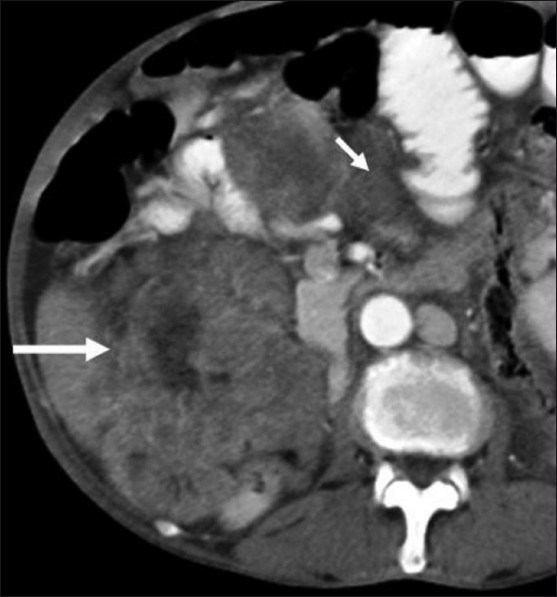
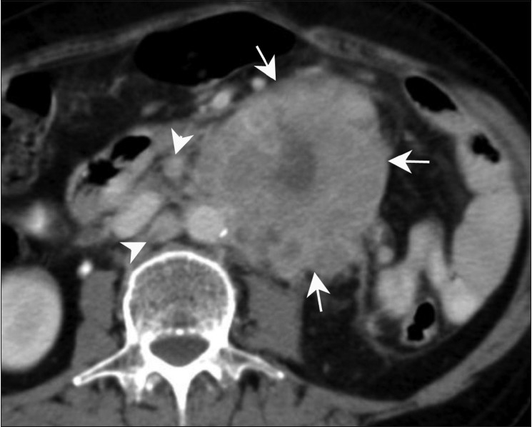
GIST with lymphadenopathy. Axial contrast-enhanced CT scan in a 52-year-old male shows a lesser sac necrotic mass (small arrow) infiltrating the pancreas with lymphadenopathy (large arrow)
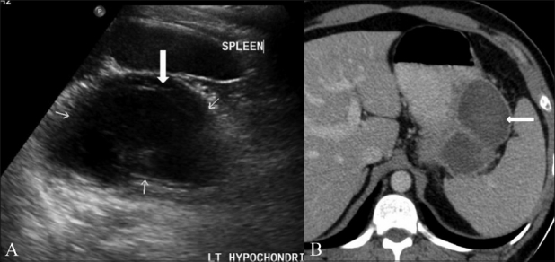
Cystic GIST. Longitudinal USG scan of the abdomen (A) in a 41-year-old male shows a predominantly cystic mass with septae (arrows) in the left hypochondrium. Axial contrast-enhanced CT scan (B) shows a peripherally enhancing cystic mass (arrow) in the lesser sac region along the tail of the pancreas
GIST with metastasis. Coronal contrast-enhanced CT scan in a 39-year-old male operated for gastric GIST shows metastasis (arrow) in the left lobe of the liver
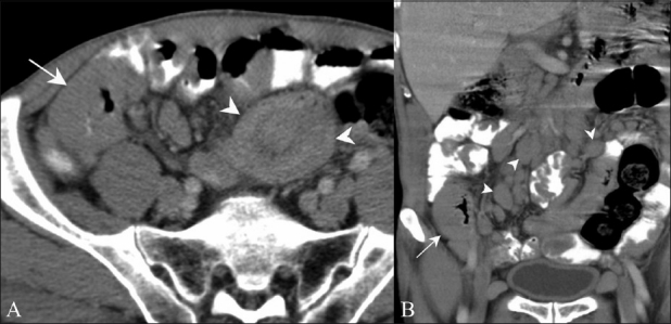
Adenocarcinoma. Axial contrast-enhanced CT scan in a 65-year-old female presenting with pain in the abdomen show a large heterogenous mass lesion (arrows) arising from the jejunal loops with para-aortic lymph nodes (arrowheads). No proximal obstruction is seen
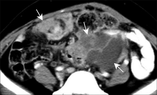
Mucosa-associated lymphoid tissue (MALT) Lymphoma. Axial contrast-enhanced CT scan (A) in a 67-year-old male presenting with generalized lymphadenopathy shows bowel wall thickening (arrow) along with intussusception (arrowhead). Coronal contrast-enhanced CT scan (B) of the same patient shows bowel wall thickening (arrow) with multiple enlarged lymph nodes (arrowheads)
Peritoneal carcinomatosis. Axial contrast-enhanced CT scan in a known case of carcinoma ovary in a middle-aged woman shows peritoneal deposits (arrows) suggestive of peritoneal carcinomatosis
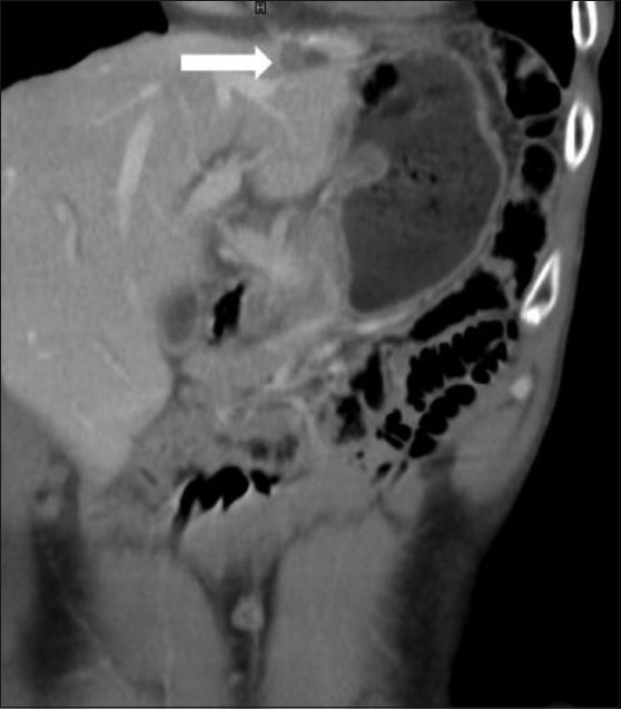
Carcinoid. Axial contrast-enhanced CT scan (A) shows a duodenal mass with intraluminal and exophytic components (arrows) along with hypervascular metastasis (arrowhead). Sagittal contrast-enhanced CT scan (B) confirms an intraluminal mass (arrow) in the duodenum causing proximal obstruction
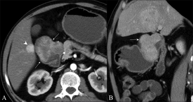
Exophytic pancreatic neuroendocrine tumor. Contrast-enhanced axial (A) and coronal (B) CT scans in a 60-year-old female show a well-defined, heterogeneously enhancing mass (arrows) along the second part of the duodenum (arrowhead)
References
-
- Burkill GJ, Badran M, Al-Muderis O, Meirion Thomas J, Judson IR, Fisher C, et al. Malignant gastrointestinal stromal tumor: Distribution, imaging features, and pattern of metastatic spread. Radiology. 2003;226:527–32. - PubMed
-
- Miettinen M, Lasota J. Gastrointestinal stromal tumors: Definition, clinical, histological, immunohistochemical, and molecular genetic features and differential diagnosis. Virchows Arch. 2001;438:1–12. - PubMed
-
- Ludwig DJ, Traverso LW. Gut stromal tumors and their clinical behavior. Am J Surg. 1997;173:390–4. - PubMed
-
- Miettinen M, Monihan JM, Sarlomo-Rikala M, Kovatich AJ, Carr NJ, Emory TS, et al. Gastrointestinal stromal tumors/smooth muscle tumors (GISTs) primary to the omentum and mesentery. Am J Surg Pathol. 1999;23:1109–18. - PubMed
-
- Fletcher CD, Berman JJ, Corless C, Gorstein F, Lasota J, Longley BJ, et al. Diagnosis of gastrointestinal stromal tumors: A consensus approach. Hum Pathol. 2002;33:459–65. - PubMed
LinkOut - more resources
Full Text Sources