

Case report: Thrombosed giant cavernous carotid artery aneurysm secondary to cervical internal carotid artery dissection: An unusual entity
- PMID: 22013300
- PMCID: PMC3190497
- DOI: 10.4103/0971-3026.85373
Case report: Thrombosed giant cavernous carotid artery aneurysm secondary to cervical internal carotid artery dissection: An unusual entity
Abstract
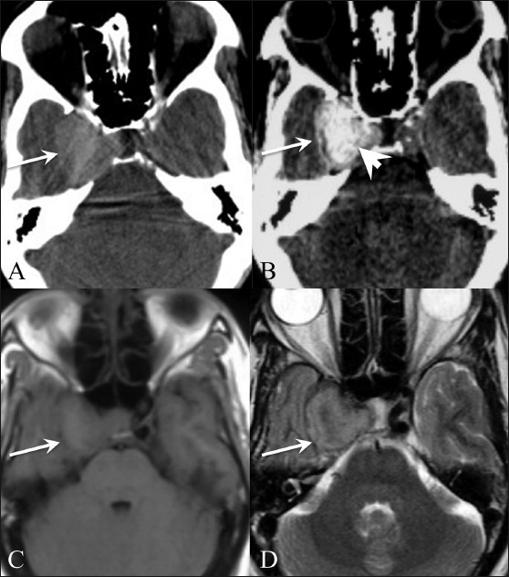
Spontaneous thrombosis of a giant intracranial aneurysm with parent artery occlusion is known. The exact mechanism is however unclear and various theories have been proposed. We present an unusual case of an angiographically documented cervical internal carotid artery (ICA) dissection, which led to total occlusion of the ICA distal to the dissected site, with acute cessation of forward blood flow. This resulted in acute upstream thrombosis of the giant cavernous carotid artery aneurysm and an acute cavernous sinus syndrome-like presentation.
Keywords: Cavernous carotid artery; dissection, giant aneurysm; internal carotid artery; thrombosis.
Conflict of interest statement
Figures

References
-
- Whittle IR, Williams DB, Halmagyi GM, Besser M. Spontaneous thrombosis of a giant intracranial aneurysm and ipsilateral internal carotid artery. J Neurosurg. 1982;56:287–9. - PubMed
-
- Kurokawa R, Kuroshima Y, Yoshida K, Kawase T. Spontaneous thrombosis of intracavernous internal carotid artery aneurysm and parent artery occlusion in patients with positive balloon test occlusion.Two case reports. Neurol Med Chir (Tokyo) 2001;41:436–41. - PubMed
-
- Kasliwal MK, Suri A, Sai Kiran NA, Sharma BS. Spontaneous Thrombosis of giant cavernous internal carotid artery aneurysm in a neonate: Case report and review of the literature. Pediatr Neurosurg. 2008;44:329–32. - PubMed
-
- Tsutsumi M, Kazekawa K, Tanaka A, Ueno Y, Nomoto Y. Spontaneous thrombosis of a giant intracavernous internal carotid artery aneurysm and ipsilateral internal carotid artery occlusion. Radiat Med. 2002;20:261–3. - PubMed
-
- Perrini P, Bortolotti C, Wang H, Fraser K, Lanzino G. Thrombosed giant intracavernous aneurysm with subsequent spontaneous ipsilateral carotid artery occlusion. Acta Neurochir (Wien) 2005;147:215–6. - PubMed
LinkOut - more resources
Full Text Sources
Miscellaneous