

Case Report: Floating fat globule within an arachnoid cyst
- PMID: 22013301
- PMCID: PMC3190498
- DOI: 10.4103/0971-3026.85374
Case Report: Floating fat globule within an arachnoid cyst
Abstract
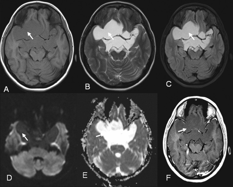
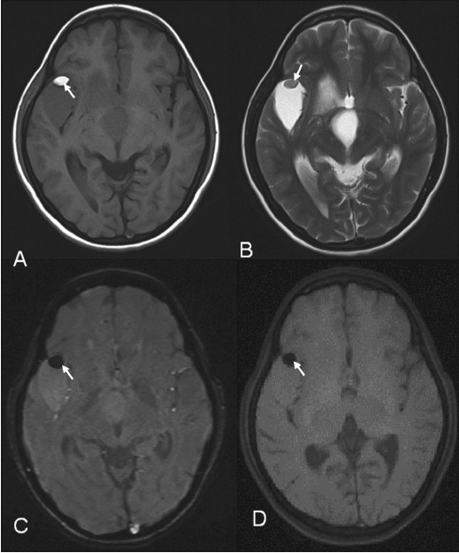
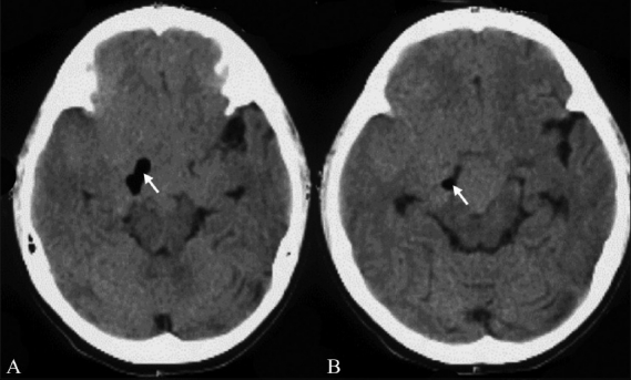
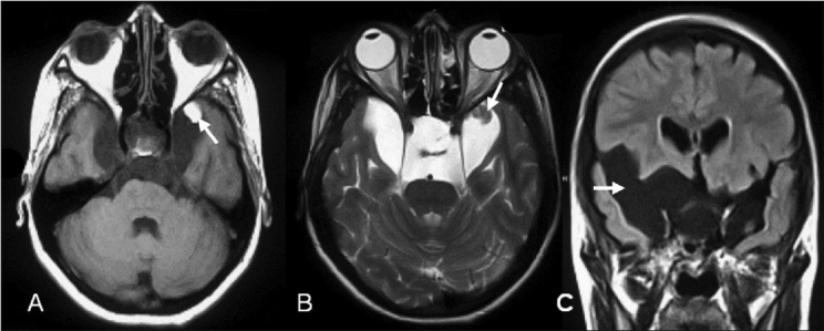
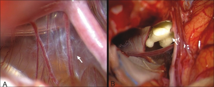
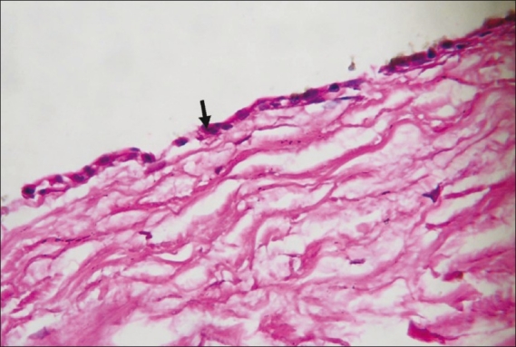
Intralesional floating fat globules have been reported in cystic lesions such as teratoma of the ovary and dermoid of the head and neck but not within intracranial lesions. Fat globules dispersed within the subarachnoid space are a known imaging finding of ruptured intracranial dermoid. We report a unique case of an intralesional solitary floating fat globule within a multicompartmental arachnoid cyst, with varying locations on serial imaging. We also put forward a hypothesis for the pathogenesis of fat within an arachnoid cyst. To the best of our knowledge, this is the first such report in the literature.
Keywords: Arachnoid cyst; intralesional fat; magnetic resonance imaging.
Conflict of interest statement
Figures
References
-
- Akyuz M, Goksu E, Aralasmak A, Tuncer R. Retroclival arachnoid cyst presenting with haemorrhage: A brief report of a special case. Acta Neurochir (Wien) 2010;152:161–2. - PubMed
-
- Burger PC, Scheithauer BW, Vogel FS. Surgical pathology of the brain and its coverings. 4th ed. Philadelphia PA: Churchill Livingstone; 2002. Intracranial meninges; pp. 89–93.
-
- McLendon RE, Tien RD. Russell and Rubinstein's pathology of tumors of the nervous system. 6th ed. New York NY: Oxford University Press; 1998. Tumors and tumorlike lesions of maldevelopmental origin; pp. 327–52.
-
- Osborn AG. Diagnostic imaging: Brain. Salt Lake City Utah: Amirsys; 2004. Arachnoid cyst; pp. I-7–4.
-
- Osborn AG. Diagnostic neuroradiology. St Louis MO: Mosby; 1994. Miscellaneous tumors, cysts, and metastases; pp. 631–49.
LinkOut - more resources
Full Text Sources