Primary spinal epidural lymphomas
- PMID: 22013369
- PMCID: PMC3190427
- DOI: 10.4103/0974-8237.85307
Primary spinal epidural lymphomas
Abstract
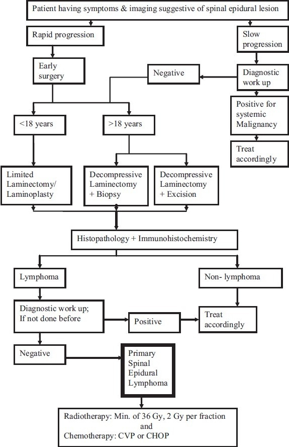
An epidural location for lymphoma is observed in 0.1-6.5% of all the lymphomas. Primary spinal epidural lymphoma (PSEL) is a subset of lymphomas, where there are no other recognizable sites of lymphomas at the time of diagnosis. The incidence of this subset of lymphomas is much less. It, however, is increasingly diagnosed, due to the increased use of more sensitive imaging modalities. For the electronic search, Pubmed was used to identify journals that enlisted and enumerated PSEL from 1961 to January 2011. The following combination of terms: "primary," "spinal," "epidural," and "lymphoma" were used. The most significant articles and their bibliographies were analyzed by the authors. The symptoms, pathogenesis, diagnostic workup, histopathology, treatment, and outcome have been analyzed in a systematic manner.
Keywords: Epidural tumors; primary spinal epidural lymphomas; spinal lymphomas.
Conflict of interest statement
Figures
References
-
- Mesfin FB, Drazin D, Berry S, Homan S, Nazeer T, German JW. Diffuse follicle center lymphoma of the spine: A primary epidural lymphoma? Clin Neuropathol. 2009;28:395–9. - PubMed
-
- Cağavi F, Kalayci M, Tekin IO, Numanoğlu G, Cağavi Z, Gül S, et al. Primary spinal extranodal Hodgkin's disease at two levels. Clin Neurol Neurosurg. 2006;108:168–73. - PubMed
-
- Ultmann JE, DeVita VT., Jr . Hodgkin's disease and other lymphomas. In: Petersdorf RG, Adams RD, Braunwald E, Isselbacher KJ, Martin JB, Wilson JD, editors. Harrison's Principles of Internal Medicine. 10th Ed. New York: McGraw-Hill; 1983. pp. 811–25.
-
- Wood NL, Coltman CA. Localized primary extranodal Hodgkin's disease. Ann Intern Med. 1973;78:113–8. - PubMed
-
- Samadian M, Vahidi S, Khormaee F, Ashraf H. Isolated, primary spinal epidural Hodgkin's disease in a child. Pediatr Neurol. 2009;40:480–2. - PubMed