Diagnostic and prognostic role of MRI in spinal trauma, its comparison and correlation with clinical profile and neurological outcome, according to ASIA impairment scale
- PMID: 22013371
- PMCID: PMC3190425
- DOI: 10.4103/0974-8237.85309
Diagnostic and prognostic role of MRI in spinal trauma, its comparison and correlation with clinical profile and neurological outcome, according to ASIA impairment scale
Abstract
Aims and objectives: To evaluate the role of magnetic resonance imaging (MRI) as a non-invasive diagnostic tool in patients with acute and chronic spinal trauma and to compare and correlate the MRI findings with those of patients' clinical profile and neurological outcome according to ASIA impairment scale to assess prognostic and clinical value of MRI.
Materials and methods: Sixty two patients of spinal trauma formed the study group in a prospective fashion. The patients undergoing MR imaging and magnetic resonance images were analyzed and correlated with findings on neurological examination according to American Spinal Injury Association (ASIA) impairment scale (AIS) at the time of MRI examination and subsequently at sub-acute interval to assess neurological outcome.
Statistical analysis: Sample profile was described in terms of 95% confidence limit and proportion. To describe strength of association between extent of spinal cord injury and outcome, odd's ratio, bivariate and multi variant analysis, was used. Pearson's chi square (χ) (2) statistics was applied to test the association between two categorical variables. Data were analyzed using statistical software package, STATA 9.2 and the difference was considered to be significant if 'P' value was <0.05.
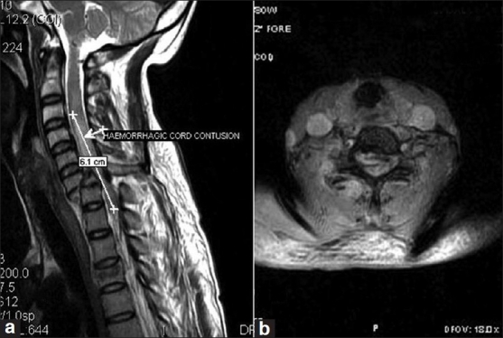
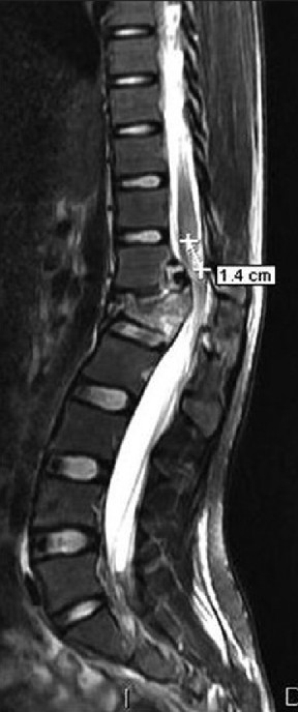
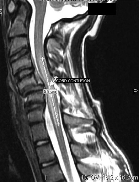
Observation and results: The cord edema without hemorrhage was the most common MR finding (41.5%). The others were sizable focus of hemorrhage within the cord (33%), epidural hematoma (5.0%), and normal cord (26%). Majority of MR findings correlated well with clinical profile of the patient according to ASIA impairment scale. This study demonstrated that patients with presence of sizable focus of haemorrhage had larger cord edema and more severe grade of initial ASIA impairment scale( AIS) with poor recovery at follow up (P=0.032).Improvement in upper extremity was more than lower extremity. Severe cord compression was also associated with poor neurological outcome; however it was not statistically significant (P=0.149).
Conclusions: With this study the authors concluded that various MRI findings in acute spinal cord injury correlated well with the initial clinical findings and on follow-up according to ASIA impairment scale. MRI is useful for initial diagnosis of acute spinal cord injury and its prognostication for predicting neurological recovery.
Keywords: ASIA impairment scale; MRI; acute spinal cord injury; prognostication; spinal trauma.
Conflict of interest statement
Figures

References
-
- Kalfas I, Wilberger J, Goldberg A, Prostko ER. Magnetic resonance imaging in acute spinal cord trauma. Neurosurgery. 1988;23:295–9. - PubMed
-
- Goldberg AL, Daffner RH, Schapiro RL. Imaging of acute spinal trauma: An evolving multi-modality approach. Clin Imag. 1990;14:11–6. - PubMed
-
- Wittenberg RH, Boetel U, Beyer HK. Magnetic resonance imaging and computed tomography of acute spinal cord trauma. Clin Orthop. 1990;260:176–85. - PubMed
-
- Flanders AE, Schaefer DM, Doan HT, Mishkin MM, Gonzalez CF, Northrup BE. Acute cervical spine trauma: Correlation of MRI imaging findings with degree of neurologic deficit. Radiology. 1990;177:25–33. - PubMed