Intramedullary enterogenous cyst of the conus medullaris presenting as lower limb pain
- PMID: 22013376
- PMCID: PMC3190431
- DOI: 10.4103/0974-8237.85314
Intramedullary enterogenous cyst of the conus medullaris presenting as lower limb pain
Abstract
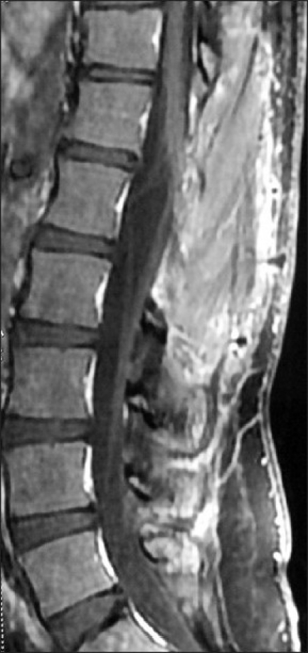
Enterogenous cysts account for 0.7-1.3% of spinal axis tumors. Cervical and thoracic segments are most often affected and they are rare in the lumbar region. Intramedullary variant which comprises less than 5% of enterogenous cysts are densely adherent to the surrounding tissue and preclude total excision. Partial excision is associated with recurrence and is the most common unfavorable outcome in these cysts. Hence, such patients need follow-up with serial imaging. We describe a case of conus medullaris enterogenous cyst presenting as lower limb pain. Due to dense adhesion of the cyst to the surrounding neural tissue, subtotal excision was done. The patient is symptom and tumor free at one year interval. We describe our case, discuss its uniqueness and review the literature on this rare but difficult to cure tumor.
Keywords: Conus medullaris tumor; enterogenous cyst; intramedullary tumor; lower limb pain; neurenteric cyst.
Conflict of interest statement
Figures


References
-
- Harriman DG. An intraspinal enterogenous cyst. J Pathol Bacteriol. 1958;75:413–9. - PubMed
-
- Fortuna A, Mercuri S. Intradural spinal cysts. Acta Neurochir (Wien) 1983;68:289–314. - PubMed
-
- D’Almeida AC, Stewart DH., Jr Neurenteric cyst. Case report and literature review. Neurosurgery. 1981;8:596–9. - PubMed
-
- Arslan E, Cakir E, Kuzeyli K, Guvercin AR, Gazioglu G, Arslan EA, et al. Recurrent lumbar spinal intradural enterogenous cyst.A Case report. Turk Neurosurg. 2010;20:402–5. - PubMed