

Cellular modulation of polymeric device surfaces: promise of adult stem cells for neuro-prosthetics
- PMID: 22013407
- PMCID: PMC3189638
- DOI: 10.3389/fnins.2011.00114
Cellular modulation of polymeric device surfaces: promise of adult stem cells for neuro-prosthetics
Abstract
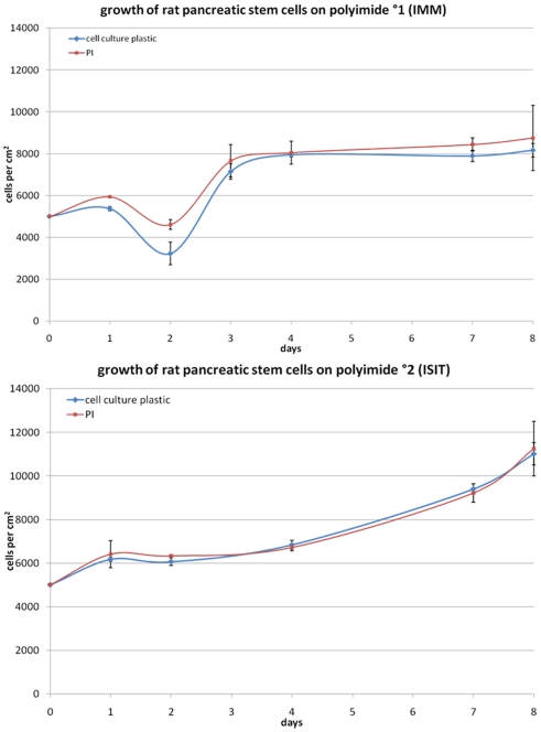
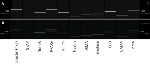
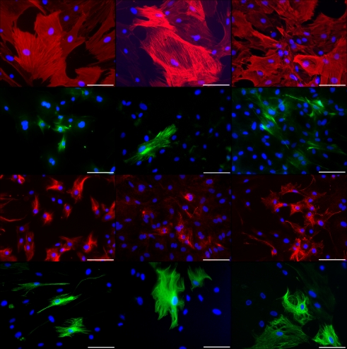
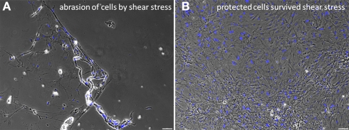
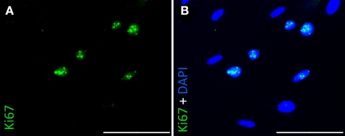
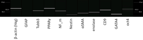
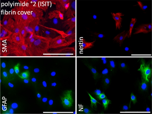
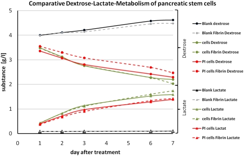
Minimizing the foreign body response is seen as one critical research strategy for implants especially when designed for immune-privileged organs like the brain. The context of this work is to improve deep brain stimulating devices used in a consistently growing spectrum of psychomotor and psychiatric diseases mainly in form of stiff electrodes. Based on the compliance match hypothesis of biocompatibility we present another step forward using flexible implant materials covered with brain cell-mimicking layers. We covered two types of flexible polyimide films with glandular stem cells derived from pancreatic acini. Using real time-PCR and fluorescent immunocytochemistry we analyzed markers representing various cell types of all three germ layers and stemness. The results demonstrate an unchanged differentiation potential of the polyimide fixated cells as measured by mRNA and protein level. Additionally we developed a fibrinous hydrogel coating to protect them against shear forces upon eventual implantation. By repeating previous analysis and additional metabolism tests for all stages we corroborate the validity of this improvement. Consequently we assume that a stem cell-containing cover may provide a native, fully and actively integrating brain-mimicking interface to the neuropil.
Keywords: fibrin; foreign body response; neural prosthesis; polyimide; stem cell; surface modification.
Figures
Similar articles
-
Ultrasoft microwire neural electrodes improve chronic tissue integration.Acta Biomater. 2017 Apr 15;53:46-58. doi: 10.1016/j.actbio.2017.02.010. Epub 2017 Feb 6. Acta Biomater. 2017. PMID: 28185910 Free PMC article.
-
Seeding neural progenitor cells on silicon-based neural probes.J Neurosurg. 2010 Sep;113(3):673-81. doi: 10.3171/2010.1.JNS09313. J Neurosurg. 2010. PMID: 20151783
-
Time course study of long-term biocompatibility and foreign body reaction to intraneural polyimide-based implants.J Biomed Mater Res A. 2018 Mar;106(3):746-757. doi: 10.1002/jbm.a.36274. Epub 2017 Nov 18. J Biomed Mater Res A. 2018. PMID: 29052368
-
Biocompatibility screening in cardiovascular implants.Z Kardiol. 2005 Jun;94(6):383-91. doi: 10.1007/s00392-005-0231-4. Z Kardiol. 2005. PMID: 15940438 Review.
-
Multifunctional Fibers as Tools for Neuroscience and Neuroengineering.Acc Chem Res. 2018 Apr 17;51(4):829-838. doi: 10.1021/acs.accounts.7b00558. Epub 2018 Mar 21. Acc Chem Res. 2018. PMID: 29561583 Review.
Cited by
-
Quantum Dots Do Not Alter the Differentiation Potential of Pancreatic Stem Cells and Are Distributed Randomly among Daughter Cells.Int J Cell Biol. 2013;2013:918242. doi: 10.1155/2013/918242. Epub 2013 Jul 24. Int J Cell Biol. 2013. PMID: 23997768 Free PMC article.
-
Bio-inspired hybrid microelectrodes: a hybrid solution to improve long-term performance of chronic intracortical implants.Front Neuroeng. 2014 Apr 10;7:7. doi: 10.3389/fneng.2014.00007. eCollection 2014. Front Neuroeng. 2014. PMID: 24782757 Free PMC article.
-
Organic electrode coatings for next-generation neural interfaces.Front Neuroeng. 2014 May 27;7:15. doi: 10.3389/fneng.2014.00015. eCollection 2014. Front Neuroeng. 2014. PMID: 24904405 Free PMC article. Review.
-
The chronic challenge-new vistas on long-term multisite contacts to the central nervous system.Front Neuroeng. 2015 Mar 18;8:3. doi: 10.3389/fneng.2015.00003. eCollection 2015. Front Neuroeng. 2015. PMID: 25852537 Free PMC article. No abstract available.
-
Future think: cautiously optimistic about brain augmentation using tissue engineering and machine interface.Front Syst Neurosci. 2015 May 19;9:72. doi: 10.3389/fnsys.2015.00072. eCollection 2015. Front Syst Neurosci. 2015. PMID: 26042003 Free PMC article. No abstract available.
References
-
- Benabid A. L., Koudsie A., Benazzouz A., Fraix V., Ashraf A., Le Bas J. F., Chabardes S., Pollak P. (2000a). Subthalamic stimulation for Parkinson’s disease. Arch. Med. Res. 31, 282–289 - PubMed
-
- Benabid A. L., Koudsie A., Pollak P., Kahane P., Chabardes S., Hirsch E., Marescaux C., Benazzouz A. (2000b). Future prospects of brain stimulation. Neurol. Res. 22, 237–246 - PubMed
-
- Berney A., Vingerhoets F. (2005). Novel brain stimulation techniques: therapeutic perspectives in psychiatry. Rev. Med. Suisse 1, 2162–2164, 2166. - PubMed
-
- Egana J. T., Danner S., Kremer M., Rapoport D. H., Lohmeyer J. A., Dye J. F., Hopfner U., Lavandero S., Kruse C., Machens H. G. (2009). The use of glandular-derived stem cells to improve vascularization in scaffold-mediated dermal regeneration. Biomaterials 30, 5918–592610.1016/j.biomaterials.2009.07.023 - DOI - PubMed
LinkOut - more resources
Full Text Sources