. 2012:2012:293625.
doi: 10.1155/2012/293625. Epub 2011 Oct 13.

Indirect effects of oral tolerance inhibit pulmonary granulomas to Schistosoma mansoni eggs

Affiliations

Indirect effects of oral tolerance inhibit pulmonary granulomas to Schistosoma mansoni eggs

Geraldo Magela Azevedo Jr et al. Clin Dev Immunol. 2012.

Abstract

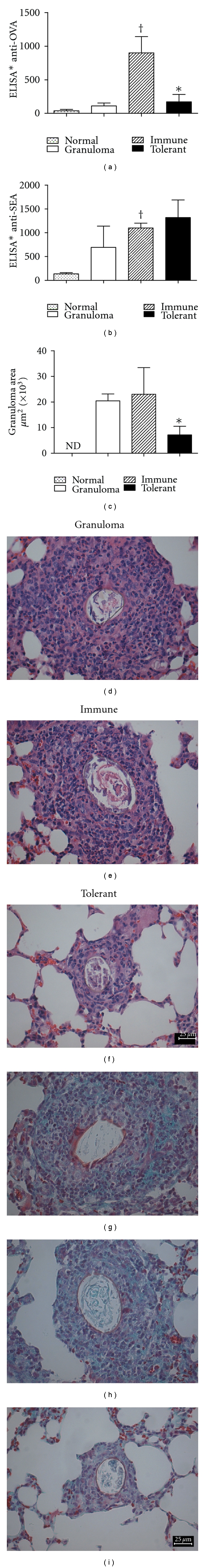
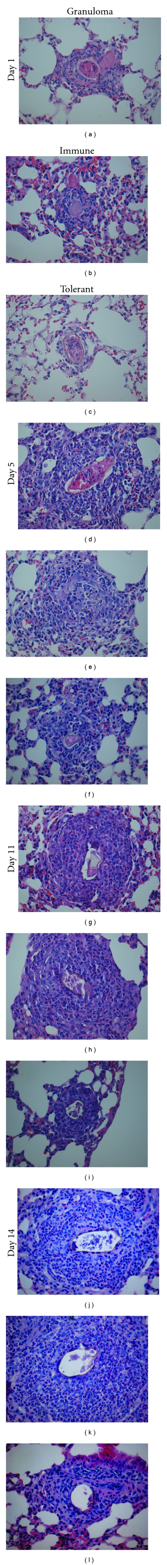
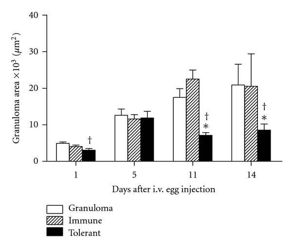
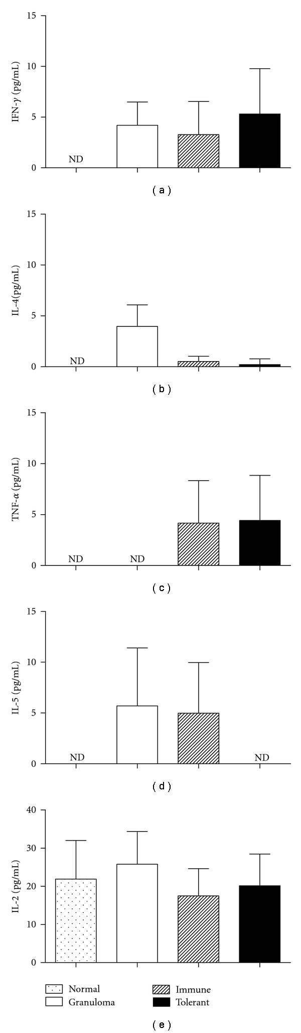
Parenteral injection of tolerated proteins into orally tolerant mice inhibits the initiation of immunological responses to unrelated proteins and blocks severe chronic inflammatory reactions of immunological origin, such as autoimmune reactions. This inhibitory effect which we have called "indirect effects of oral tolerance" is also known as "bystander suppression." Herein, we show that i.p. injection of OVA + Al(OH)(3) minutes before i.v. injection of Schistosoma mansoni eggs into OVA tolerant mice blocked the increase of pulmonary granulomas. In addition, the expression of ICAM-1 in lung parenchyma in areas outside the granulomas of OVA-orally tolerant mice was significantly reduced. However, at day 18 after granuloma induction there was no difference in immunofluorescency intensity to CD3, CD4, F4/80, andα-SMA per granuloma area of tolerant and control groups. Reduction of granulomas by reexposure to orally tolerated proteins was not correlated with a shift in Th-1/Th-2 cytokines in serum or lung tissue extract.


Figures

Figure 1
Figure 1
Reduction of granuloma by re-exposure of orally tolerant animals to the tolerated antigen. Serum levels of (a) anti-OVA antibodies and (b) anti-SEA antibodies and (c) pulmonary granuloma area and (d–i) histological aspect of pulmonary granuloma 18 days after i.v. injection of S. mansoni eggs in nonimmunized mice (granuloma group, open bars), OVA immune controls (hatched bars), and OVA-orally tolerant (black bars). Normal mice (doted bars) were not immunized with OVA neither injected with eggs. Data represent mean ± SEM. *P ≤ 0.05 tolerant versus immune P ≤ 0.05 immune versus normal. nd: not detected. Original magnification of HE (d–f) and Gomori's trichrome (g–i) photomicrographs 400X; scale bars = 25 μm.
Figure 2
Figure 2
Granuloma at different times after egg injection. Lung HE staining 1, 5, 11, and 14 days after i.v. injection of S. mansoni eggs. (a–c) At day 1, an inflammatory infiltrate with predominance of neutrophils and macrophages can be detected around eggs in all groups, but it is less intense in the tolerant group. (d–f) At day 5, macrophages, eosinophils, and some lymphocytes can be detected. (g–l) At days 11 and 14 granulomas are more organized and some fibroblasts can be detected. Granulomas in tolerant mice follow the same pattern of organization but do not reach the same size of granulomas in controls group. Scale bars = 25 μm.
Figure 3
Figure 3
Re-exposure of orally tolerant animals to the tolerated antigen block enlargement of granuloma area. The area of granulomas at days 1, 5, 11 and 14 after i.v. injection of eggs in nonimmunized mice (granuloma group, open bars), OVA immune controls (hatched bars), and OVA-orally tolerant (black bars). Data represent mean ± SEM (five mice/group). *P ≤ 0.05 tolerant versus immune and P ≤ 0.05 tolerant versus granuloma.
Figure 4
Figure 4
Cell subsets in pulmonary granulomas 18 days after i.v. egg injection. Immunolocalization using specific antibodies followed by secondary antibodies coupled with fluorescein (green) and nuclear counterstainig with 4′6-diamidino-2-phenylindol (blue), 18 days after i.v. eggs injection. Confocal microscope images were captured with a 63X objective, and the graphs represent the green fluorescence intensity (the sum of gray values of all pixels divided by the area (in μm2) × 10−3) of expression of F4/80 (macrophages), CD3 (T-lymphocytes), CD4+ cells, and α-SMA (myofibroblasts) in nonimmunized mice (granuloma group, open bars), OVA immune controls (hatched bars), and OVA-orally tolerant (black bars). Data represent mean ± SEM of fluorescence intensity of duplicate slides (n = 5 mice/group). The green autofluorescency of eggs was excluded from all analyses. No significant difference was found between groups.
Figure 5
Figure 5
Re-exposure of orally tolerant animals to the tolerated antigen block the rise of ICAM-1 expression in lung parenchyma. Immunolocalization of ICAM-1 in granulomas and lung parenchyma using specific antibody coupled with fluorescein (green) and nuclear counterstainig with 4′6-diamidino-2-phenylindol (blue), 18 days after i.v. eggs injection. Confocal microscope images were captured with a 63X objective, and the graphs represent the green fluorescence intensity (the sum of gray values of all pixels divided by the area (in μm2) × 10−3) of expression of ICAM-1 in the granuloma area (a–d) and in lung parenchyma (e–h) in nonimmunized mice (granuloma group, open bars), OVA immune controls (hatched bars), and OVA-orally tolerant mice (black bars). Data represent mean ± SEM of fluorescence intensity of duplicate slides (n = 5 mice/group).
Figure 6
Figure 6
Cytokines production of lungs. Fourteen days after i.v. injection of S. mansoni eggs in nonimmunized mice (granuloma group, open bars), OVA immune controls (hatched bars), and OVA-orally tolerant mice (black bars), lungs were removed and homogenized in extract buffer. Normal mice (doted bars) were not immunized with OVA and neither injected with eggs. Extract supernatant was collected for cytokine assay. IFN-γ, TNF-α, IL-2, IL-4, and IL-5 were measured using a Cytometric Bead Array (CBA) kit. The results are shown as mean concentrations ± SEM. nd: not detected.
Figure 7
Figure 7
Time course of serum cytokines after granuloma induction. IFN-γ, TNF-α, IL-2, IL-4, and IL-5 were measured using a Cytometric Bead Array (CBA) kit in serum samples collected from nonimmunized mice (granuloma group, open bars), OVA immune controls (hatched bars), and OVA-orally tolerant mice (black bars). Normal mice (doted bars) were not immunized with OVA and neither injected with eggs. The results are shown as mean concentrations ± SEM. nd: not detected. IL-2 and IL-4 were not detected.
Figure 8
Figure 8
IFN-γ and IL-10 production of spleen cells stimulated with SEA or OVA. Eighteen days after i.v. injection of S. mansoni eggs in nonimmunized mice (granuloma group, open bars), OVA immune controls (hatched bars), and OVA-orally tolerant mice (black bars) spleen cells were cultured with medium, ConA, SEA or OVA for 3 days. Normal mice (doted bars) were not immunized with OVA neither injected with eggs. The culture supernatant fluids were harvested and IFN-γ and IL-10 measured by sandwich ELISA. The results are shown as mean concentrations ± SEM. nd: not detected.

Similar articles

Cited by

References

    1. Faria AM, Weiner HL. Oral tolerance: therapeutic implications for autoimmune diseases. Clinical and Developmental Immunology. 2006;13(2–4):143–157. - PMC - PubMed
    1. Vaz N, Faria AM, Verdolin BA, Carvalho CR. Immaturity, ageing and oral tolerance. Scandinavian Journal of Immunology. 1997;46(3):225–229. - PubMed
    1. Miller A, Lider O, Weiner H. Antigen-driven bystander suppression after oral administration of antigens. Journal of Experimental Medicine. 1991;174(4):791–798. - PMC - PubMed
    1. Russo M, Jancar S, Pereira de Siqueira AL, et al. Prevention of lung eosinophilic inflammation by oral tolerance. Immunology Letters. 1998;61(1):15–23. - PubMed
    1. Yoshino S, Quattrocchi E, Weiner HL. Suppression of antigen-induced arthritis in Lewis rats by oral administration of type II collagen. Arthritis and Rheumatism. 1995;38(8):1092–1096. - PubMed

Publication types

MeSH terms