. 2011 Nov 29;50(47):10231-40.
doi: 10.1021/bi200801z. Epub 2011 Nov 3.

Crystal structure and characterization of particulate methane monooxygenase from Methylocystis species strain M

Affiliations

Crystal structure and characterization of particulate methane monooxygenase from Methylocystis species strain M

Stephen M Smith et al. Biochemistry. .

Abstract

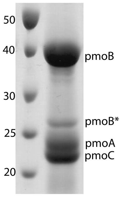
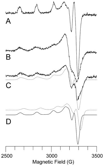
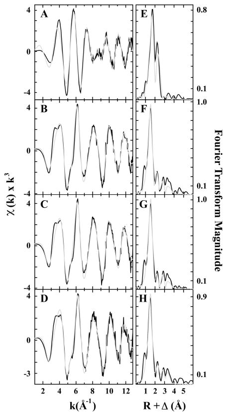
Particulate methane monooxygenase (pMMO) is an integral membrane metalloenzyme that oxidizes methane to methanol in methanotrophic bacteria. Previous biochemical and structural studies of pMMO have focused on preparations from Methylococcus capsulatus (Bath) and Methylosinus trichosporium OB3b. A pMMO from a third organism, Methylocystis species strain M, has been isolated and characterized. Both membrane-bound and solubilized Methylocystis sp. strain M pMMO contain ~2 copper ions per 100 kDa protomer and exhibit copper-dependent propylene epoxidation activity. Spectroscopic data indicate that Methylocystis sp. strain M pMMO contains a mixture of Cu(I) and Cu(II), of which the latter exhibits two distinct type 2 Cu(II) electron paramagnetic resonance (EPR) signals. Extended X-ray absorption fine structure (EXAFS) data are best fit with a mixture of Cu-O/N and Cu-Cu ligand environments with a Cu-Cu interaction at 2.52-2.64 Å. The crystal structure of Methylocystis sp. strain M pMMO was determined to 2.68 Å resolution and is the best quality pMMO structure obtained to date. It provides a revised model for the pmoA and pmoC subunits and has led to an improved model of M. capsulatus (Bath) pMMO. In these new structures, the intramembrane zinc/copper binding site has a different coordination environment from that in previous models.


Figures

Figure 1
Figure 1
SDS-PAGE gel of solubilized Methylocystis sp. strain M pMMO. Molecular mass standards are labeled in kDa. The band labeled pmoB* corresponds to a proteolytic fragment of pmoB with an N-terminus starting at residue Gln 260.
Figure 2
Figure 2
X-band EPR spectra of Methylocystis sp. strain M pMMO. (A) Cu-EDTA standard (250 μM). (B) Membrane-bound pMMO. (C) Solubilized pMMO with simulation (dashed line) generated by summing two contributing type 2 CuII centers. (D) Individual simulated spectra of the two components in (C). The experimental spectra are offset for clarity, but their intensities are meaningful since the spectra were recorded under identical conditions. Experimental parameters: microwave frequency, 9.21 GHz; microwave power, 15 dB; modulation amplitude, 5.000 G; receiver gain, 1.25 × 10²; time constant, 0.3 s; temperature, 77 K; 2 min scan. Simulation parameters for major component (solid line in part D): relative integrated intensity, 1.0; g = 2.052, g|| = 2.247, A(63Cu)|| = 570 MHz, A (63Cu) = 60 MHz. Simulation parameters for minor component (dashed line in part D): relative integrated intensity, 0.7; g = 2.04, g|| = 2.215, A(63Cu)|| = 570 MHz, A(63Cu) = 60 MHz.
Figure 3
Figure 3
Copper XANES spectra of Methylocystis sp. strain M pMMO. The spectra for (A) reduced, (B) as-isolated, (C) Cu-reconstituted, membrane-bound, and (D) oxidized samples are shown. The reduced, as-isolated, and oxidized samples were solubilized. The dashed vertical line at ca. 8984 eV identifies spectral features corresponding to the CuI 1s → 4p transition. Spectra are offset for clarity.
Figure 4
Figure 4
Copper EXAFS fitting analysis for Methylocystis sp. strain M pMMO. Raw EXAFS data for (A) reduced, (B) as-isolated, (C) Cu-reconstituted, membrane-bound, and (D) oxidized pMMO are shown in black. The reduced, as-isolated, and oxidized samples were solubilized. The phase shifted Fourier transforms of the copper data for (E) reduced, (F) as-isolated, (G) Cu-reconstituted, membrane-bound, and (H) oxidized are shown in black. Best-fit simulations are shown in gray.
Figure 5
Figure 5
Structures of Methylocystis sp. strain M pMMO subunits. (A) One of the three protomers in the trimer. The pmoB, pmoA, and pmoC subunits are shown in gray, blue, and purple, respectively. Copper ions are shown in cyan, and a zinc ion is shown in gray. The additional helix is shown in yellow. (B) Superposition of the pmoA subunits from the Methylocystis sp. strain M (blue) and the original M. capsulatus (Bath) (gray) structures. (C) Superposition of the pmoC subunits from the Methylocystis sp. strain M (purple) and the original M. capsulatus (Bath) (light pink) structures. Residues discussed in the text are labeled.
Figure 6
Figure 6
Metal centers in Methylocystis sp. strain M pMMO. The copper sites in (A) and (B) were modeled as dinuclear and mononuclear, respectively. The Cu anomalous map (magenta, 5σ) is superposed on the 2FoFc map (1.4σ). (C) The zinc site is solvent exposed. The Zn anomalous map (cyan, 4.5σ) is superposed on the 2FoFc map (1.4σ). (D) The zinc site as modeled in the original M. capsulatus (Bath) pMMO structure. The pmoC and pmoA subunits are colored light pink and gray as in Figure 5.

Similar articles

Cited by

References

    1. Hanson RS, Hanson TE. Methanotrophic bacteria. Microbiol Rev. 1996;60:439–471. - PMC - PubMed
    1. Hakemian AS, Rosenzweig AC. The biochemistry of methane oxidation. Annu Rev Biochem. 2007;76:223–241. - PubMed
    1. Rosenzweig AC, Frederick CA, Lippard SJ, Nordlund P. Crystal structure of a bacterial non-haem iron hydroxylase that catalyses the biological oxidation of methane. Nature. 1993;366:537–543. - PubMed
    1. Merkx M, Kopp DA, Sazinsky MH, Blazyk JL, Müller J, Lippard SJ. Dioxygen activation and methane hydroxylation by soluble methane monooxygenase: a tale of two irons and three proteins. Angew Chem, Int Ed. 2001;40:2782–2807. - PubMed
    1. Tinberg CE, Lippard SJ. Dioxygen activation in soluble methane monooxygenase. Acc Chem Res. 2011;44:280–288. - PMC - PubMed

Publication types

MeSH terms

LinkOut - more resources