. 2012 May;166(2):557-68.
doi: 10.1111/j.1476-5381.2011.01721.x.

Blockade of I(Ca) suppresses early afterdepolarizations and reduces transmural dispersion of repolarization in a whole heart model of chronic heart failure

Affiliations

Blockade of I(Ca) suppresses early afterdepolarizations and reduces transmural dispersion of repolarization in a whole heart model of chronic heart failure

P Milberg et al. Br J Pharmacol. 2012 May.

Abstract

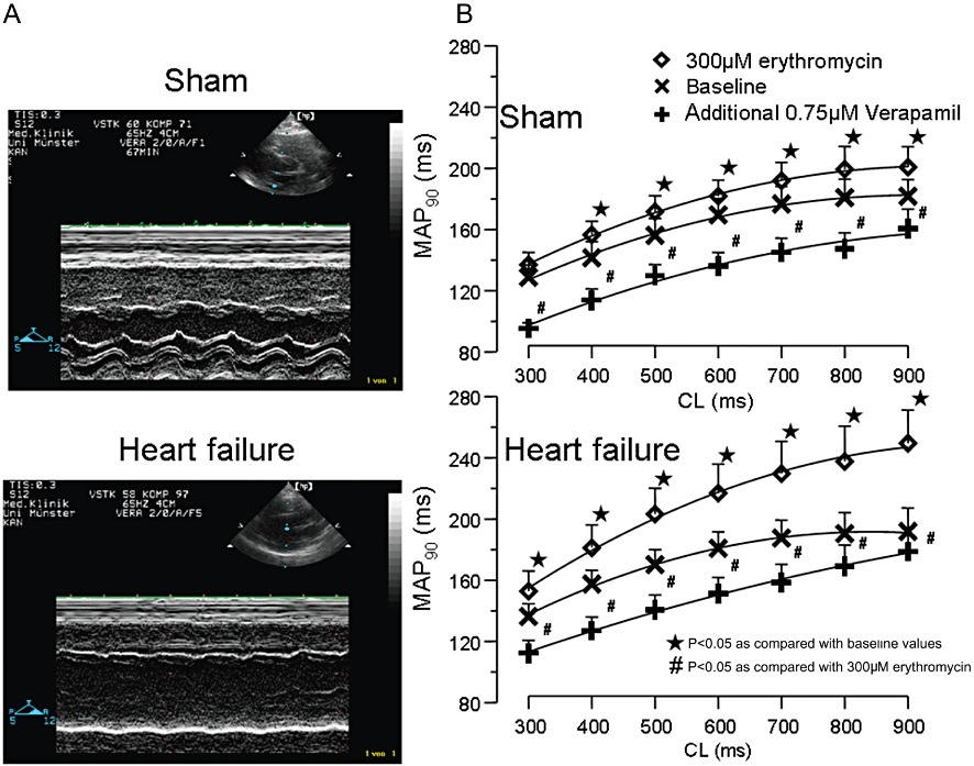
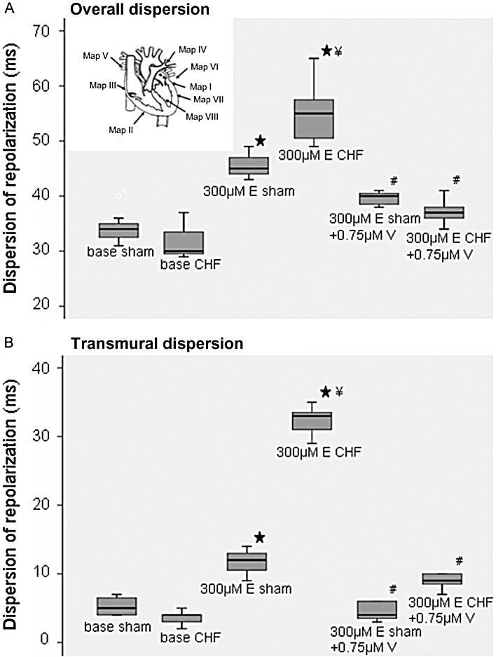
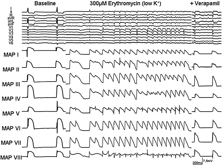
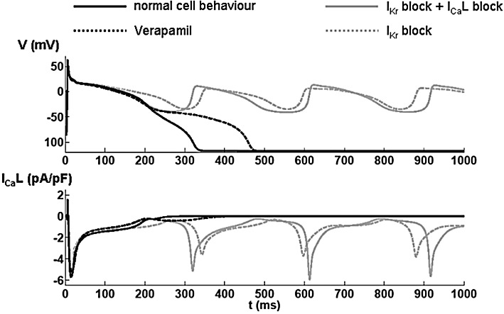
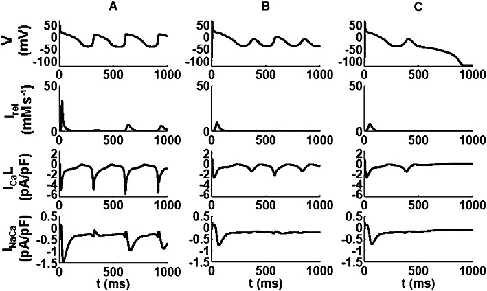
BACKGROUND AND PURPOSE Chronic heart failure (CHF) is associated with action potential prolongation and Ca(2+) overload, increasing risk of ventricular tachyarrhythmias (VT). We therefore investigated whether I(Ca) blockade was anti-arrhythmic in an intact perfused heart model of CHF. EXPERIMENTAL APPROACH CHF was induced in rabbits after 4 weeks of rapid ventricular pacing. Hearts from CHF and sham-operated rabbits were isolated and perfused (Langendorff preparation), with ablation of the AV node. VT was induced by erythromycin and low [K(+) ] (1.5mM). Electrophysiology of cardiac myocytes, with block of cation currents, was simulated by a mathematical model. KEY RESULTS Repolarization was prolonged in CHF hearts compared with sham-operated hearts. Action potential duration (APD) and overall dispersion of repolarization were further increased by erythromycin (300 µM) to block I(Kr) in CHF hearts. After lowering [K(+) ] to 1.5mM, CHF and sham hearts showed spontaneous episodes of polymorphic non-sustained VT. Additional infusion of verapamil (0.75 µM) suppressed early afterdepolarizations (EAD) and VT in 75% of sham and CHF hearts. Verapamil shortened APD and dispersion of repolarization, mainly by reducing transmural dispersion of repolarization via shortening of endocardial action potentials. Mathematical simulations showed that EADs were more effectively reduced by verapamil assuming a state-dependent block than a simple block of I(Ca) . CONCLUSIONS AND IMPLICATIONS Blockade of I(Ca) was highly effective in suppressing VT via reduction of transmural dispersion of repolarization and suppression of EAD. Such blockade might represent a novel therapeutic option to reduce risk of VT in structurally normal hearts and also in heart failure. LINKED ARTICLE This article is commented on by Stams et al., pp. 554-556 of this issue. To view this commentary visit http://dx.doi.org/10.1111/j.1476-5381.2011.01818.x.


Figures

Figure 1
Figure 1
(A) Echocardiography before Langendorff experiment in sham hearts (top) and after 4 weeks of rapid ventricular pacing (bottom). (B) Cycle length (CL)-dependent effects on action potential duration (mean MAP90) under baseline conditions, after infusion of 300 µM erythromycin and after additional infusion of 0.75 µM verapamil. ⋆P < 0.05, significantly different from baseline values; #P < 0.05, significantly different from 300 µM erythromycin).
Figure 2
Figure 2
(A) Dispersion of repolarization after erythromycin (E) and additional verapamil (V) administration in sham and heart failure (CHF) hearts (⋆P < 0.05, significantly different from baseline; ¥P < 0.05, significantly different from erythromycin treated sham hearts) and significant decrease after additional infusion of verapamil (#P < 0.01, significantly different from erythromycin-treated hearts; left top, distribution of MAP catheters). (B) Transmural dispersion of repolarization after erythromycin and additional verapamil administration (⋆P < 0.05, significantly different from baseline; ¥P < 0.05, significantly different from erythromycin-treated sham hearts; #P < 0.01, significantly different from erythromycin-treated hearts).
Figure 3
Figure 3
(A) VT incidence in sham and failing hearts after infusion of erythromycin (⋆P < 0.05, significantly different from baseline) and additional infusion of verapamil (#P < 0.05, significantly different from erythromycin treatment). (B) Number of single VT episodes in sham and failing hearts after infusion of erythromycin (⋆P < 0.05, significantly different from baseline) and additional infusion of verapamil (#P < 0.05, significantly different from erythromycin treatment).
Figure 4
Figure 4
Representative example of EAD and polymorphic VT in the presence of erythromycin and after additional infusion of verapamil during bradycardia (AV block) and hypokalaemia in an isolated Langendorff-perfused heart from a CHF rabbit. ECG characteristics and MAP recordings [distribution of MAP catheters. Left heart: MAP I = base anterior, MAP IV = base posterior, MAP V = between basis and apex (posterolateral), MAP VI = between basis and apex (inferior); MAP VII = apex, MAP VIII = endocardial; right heart: MAP II = apex, MAP III = bases].
Figure 5
Figure 5
Results from a mathematical model of the electrophysiology of single cardiac myocytes. The upper panel shows the simulated membrane potential (V) and the lower panel shows the modelled Ca2+ current through the L-type Ca2+ channels (ICaL). Each trace represents the first action potentials after a switch to hypokalaemic ([K+] 1.5 mM) conditions in the model. The solid black line represents changes in membrane potential and ICa under normal conditions. In the presence of a substantial block of IKr, EADs appear when switching to hypokalaemic conditions, as shown by the dashed grey line. When adding a simple ICa pore blocker to the IKr block, the action potential repolarization is faster, but due to reactivation of the L-type Ca2+ channels, EADs are still elicited (grey solid line). However, assuming a state-dependent block (for verapamil) substantially inhibits the reactivation and therefore only leads to a prolongation of the action potential (black dashed line).
Figure 6
Figure 6
The panels in columns A, B and C show simulations of the effects of combining substantial IKr block together with ICa block on currents through the Na/Ca exchanger (INaCa), through L-type Ca2+ channels (ICaL) and the ryanodine release current (Irel), as well as the membrane potential (V) under conditions modelling (A) sham-operated hearts and (B and C) CHF hearts. Traces in A and B show results from a simple pore-blocker, whereas those in C denote the results for the state/voltage-dependent ICa block by verapamil. The reduction in Ca2+ cycling in the failing hearts is apparent as well as the benefit of verapamil state-dependent block over the other blocking type. Note the difference in the ICaL dynamics during the EAD between the three traces.

Similar articles

Cited by

References

    1. Akar FG, Rosenbaum DS. Transmural electrophysiological heterogeneities underlying arrhythmogenesis in heart failure. Circ Res. 2003;93:638–645. - PubMed
    1. Alexander SPH, Mathie A, Peters JA. Guide to Receptors and Channels (GRAC), 5th Edition. Br J Pharmacol. 2011;164(Suppl. 1):S1–S324. - PMC - PubMed
    1. Antzelevitch C, Sun ZQ, Zhang ZQ, Yan GX. Cellular and ionic mechanisms underlying erythromycin-induced long QT intervals and torsade de pointes. J Am Coll Cardiol. 1996;28:1836–1848. - PubMed
    1. Barr CS, Naas A, Freeman M, Lang CC, Struthers AD. QT dispersion and sudden unexpected death in chronic heart failure. Lancet. 1994;343:327–329. - PubMed
    1. Bonnar CE, Davie AP, Caruana L, Fenn L, Ogston SA, McMurray JJ, et al. QT dispersion in patients with chronic heart failure: beta blockers are associated with a reduction in QT dispersion. Heart. 1999;81:297–302. - PMC - PubMed

MeSH terms