. 2012 Apr;165(8):2749-57.
doi: 10.1111/j.1476-5381.2011.01728.x.

S(+)amphetamine induces a persistent leak in the human dopamine transporter: molecular stent hypothesis

Affiliations

S(+)amphetamine induces a persistent leak in the human dopamine transporter: molecular stent hypothesis

Aldo A Rodriguez-Menchaca et al. Br J Pharmacol. 2012 Apr.

Abstract

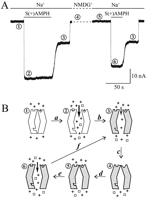
Background and purpose: Wherever they are located, dopamine transporters (DATs) clear dopamine (DA) from the extracellular milieu to help regulate dopaminergic signalling. Exposure to amphetamine (AMPH) increases extracellular DA in the synaptic cleft, which has been ascribed to DAT reverse transport. Increased extracellular DA prolongs postsynaptic activity and reinforces abuse and hedonic behaviour.

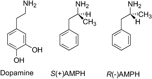
Experimental approach: Xenopus laevis oocytes expressing human (h) DAT were voltage-clamped and exposed to DA, R(-)AMPH, or S(+)AMPH.

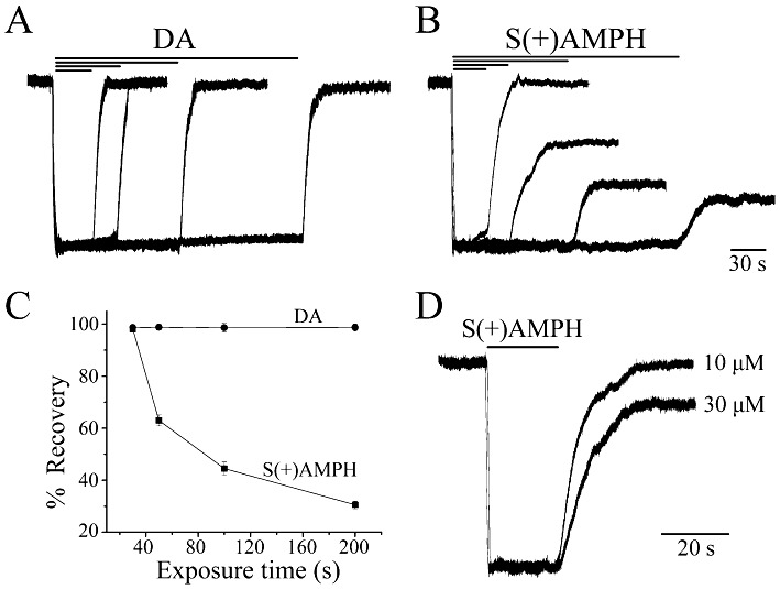
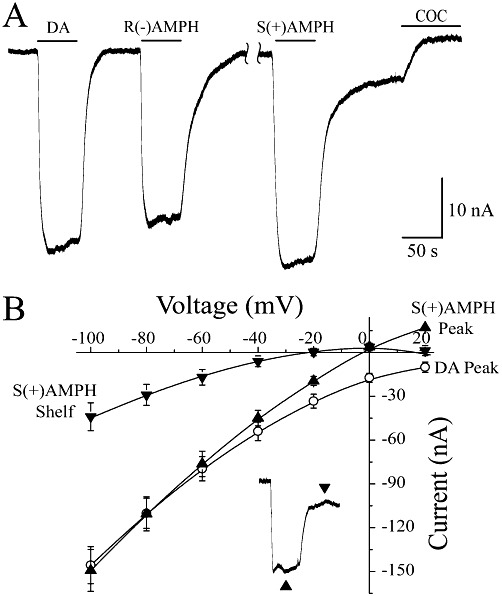
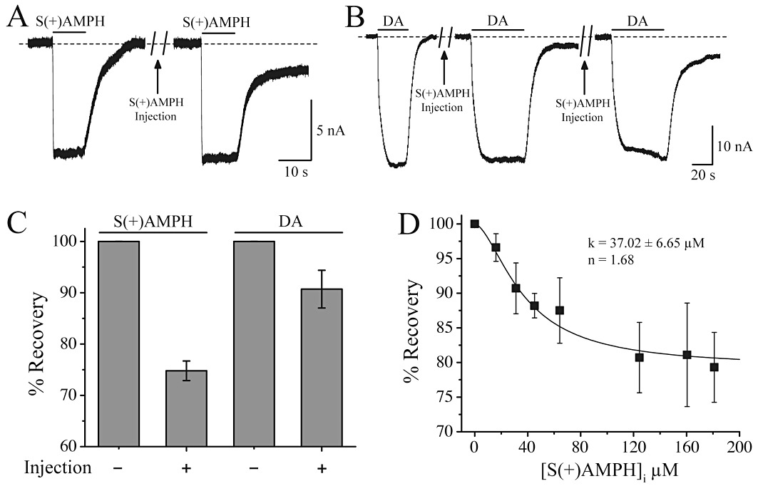
Key results: At -60mV, near neuronal resting potentials, S(+)AMPH induced a depolarizing current through hDAT, which after removing the drug, persisted for more than 30 min. This persistent leak in the absence of S(+)AMPH was in contrast to the currents induced by R(-)AMPH and DA, which returned to baseline immediately after their removal. Our data suggest that S(+)AMPH and Na(+) carry the initial S(+)AMPH-induced current, whereas Na+ and Cl(-) carry the persistent leak current. We propose that the persistent current results from the internal action of S(+)AMPH on hDAT because the temporal effect was consistent with S(+)AMPH influx, and intracellular S(+)AMPH activated the effect. The persistent current was dependent on Na(+) and was blocked by cocaine. Intracellular injection of S(+)AMPH also activated a DA-induced persistent leak current.

Conclusions and implications: We report a hitherto unknown action of S(+)AMPH on hDAT that potentially affects AMPH-induced DA release. We propose that internal S(+)AMPH acts as a molecular stent that holds the transporter open even after external S(+)AMPH is removed. Amphetamine-induced persistent leak currents are likely to influence dopaminergic signalling, DA release mechanisms, and amphetamine abuse.


Figures

Figure 1
Figure 1
Structures of dopamine and enantiomers of amphetamine. Chemical composition of dopamine, S(+)amphetamine and R(–) amphetamine (labelled S(+)AMPH and R(–)AMPH, respectively), showing their structural similarity.
Figure 2
Figure 2
S(+)AMPH induces a persistent ‘shelf’ current through the human dopamine transporter (hDAT). (A) External DA (10 µM) induced a large inward ‘peak’ current at V =−60 mV that returned to baseline when DA was removed, regardless of DA exposure time. DA peak currents were normalized to the briefest exposure time. (B) S(+)AMPH (10 µM) induced a similar inward peak current for exposures less than 30 s; however, for longer exposure times, a current that we term ‘leak’ or ‘shelf’ remained long after S(+)AMPH had been removed, and the amplitude of the shelf was proportional to the length of exposure. S(+)AMPH peak currents were normalized to the briefest exposure time. (C) Amplitude of the shelf current relative to the initial peak current plotted against exposure time of external S(+)AMPH, compared with the corresponding DA currents (n= 4, ±SEM). (D) A relatively brief and initial exposure to S(+)AMPH (20 s.), which ordinarily would not produce a shelf current, did so if the concentration of S(+)AMPH was increased from 10 to 30 µM. For the same exposure range of times and concentrations, neither DA nor S(+)AMPH induced peak or shelf currents in mock-injected oocytes (data not shown).
Figure 3
Figure 3
DA- and AMPH-induced currents. (A) At −60 mV, neither 10 µM DA nor 10 µM R(–)AMPH induced a shelf but the current always return to baseline after their removal. S(+)AMPH on the other hand induced a prominent shelf current that was blocked by cocaine (10 µM). The peak currents are approximately the same at −60 mV for 10 µM DA, R(–)AMPH, or S(+)AMPH. Note that in the presence of 10 µM cocaine the current returned to values positive to the initial baseline, indicating the presence of an endogenous leak current. (B) Baseline subtracted I(V) curves for DA, S(+)AMPH peak and S(+)AMPH shelf. The S(+)AMPH peak I(V) was shifted to the left above −60 mV compared with the DA peak, consistent with the conductance of both Na+ and S(+)AMPH through hDAT. The S(+)AMPH shelf I(V) was further shifted to the left, consistent with the absence of S(+)AMPH and presence of Cl- ions flowing through hDAT.
Figure 4
Figure 4
S(+)AMPH injection promotes the shelf current. (A) A brief application (10 s) of 10 µM external S(+)AMPH did not illicit a shelf current; however, after injecting sufficient S(+)AMPH into the cell to elevate the internal concentration to 25 µM (see Methods), the same challenge induced a prominent shelf current. (B) Dopamine, which ordinarily would not produce a shelf current (left trace), now induced a shelf current after its removal in added after a similar 25 µM injection of S(+)AMPH (centre trace). Doubling the internal S(+)AMPH concentration resulted in the generation of a larger shelf (right trace). Injecting DA into the oocyte had no similar effect on the responses to external S(+)AMPH or DA (data not shown). (C) For the same external and internal concentrations, S(+)AMPH generated a stronger shelf current than DA. Following the same protocol as in (B), we titrated internal S(+)AMPH by repeated injections into the same oocyte or single more concentrated injections in different oocytes (see Methods). (D) Pooled data for 10 µM external DA applied for 10 s: the greater the internal S(+)AMPH concentration the greater the DA-induced shelf current, that is, in the presence of internal S(+)AMPH, less of the DA-induced current was able to return to baseline after external DA removal. For a DA challenge, the shelf current saturates at 80% full recovery, with a Hill coefficient nH= 1.7 and k1/2= 37 µM.
Figure 5
Figure 5
Model for S(+)AMPH-induced, hDAT-mediated peak and shelf currents. (A) Phase 1: the baseline current at V =−60 mV in external physiological solution containing Na+. Phase 2: the 10 µM S(+)AMPH-induced peak current in Na+, nominally between 10 and 25 nA. Phase 3: the shelf current (persistent leak current), which remains after removing external S(+)AMPH. At fixed voltage, the shelf amplitude depends on exposure time and concentration of external S(+)AMPH. Phase 4: replacing Na+ with NMDG introduces a shift in baseline that we removed from the figure (dashed line); however, re-introducing Na+ during phase 4 (phase 5) does not restore the shelf current but returns the current to the original baseline [S(+)AMPH has been transported into the cell during phase 2]. Phase 6: reintroducing external S(+)AMPH in the presence of Na+ induces a new peak current. Note that the second application of S(+)AMPH is normally too brief to elicit a shelf current; however, S(+)AMPH is already present inside the cell from the first application. The new peak is smaller than the first peak, possibly due to hDAT internalization. Finally, after removing S(+)AMPH for the second time, the shelf current again manifests itself. (B) The ‘+’ symbols stand for Na+ ions, and the ‘open squares’ stand for S(+)AMPH) or in some experiments DA. The hatched transporter indicates internal occupancy by S(+)AMPH and a long-lasting ‘molecular stent’ configuration. The numbers above each state of the transporter correspond to the traces in (A). Transition (a) opens the top and bottom gates, which for brief external S(+)AMPH exposures would close. Transition (b) occurs after longer exposures and S(+)AMPH has built up inside to the extent that the inner S(+)AMPH site remains occupied and holds both gates open (molecular stent hypothesis), even in the absence of external S(+)AMPH. Transition (c): removing external Na+ closes the outer gate, which does not reopen without external Na+ and S(+)AMPH both present (transitions d and e), rendering the transporter capable of (and indeed more prone to) forming the molecular stent (transition f), because internal S(+)AMPH is still present.

Similar articles

Cited by

References

    1. Amara SG, Sonders MS. Neurotransmitter transporters as molecular targets for addictive drugs. Drug Alcohol Depend. 1998;51:87–96. - PubMed
    1. Burnette WB, Bailey MD, Kukoyi S, Blakely RD, Trowbridge CG, Justice JB., Jr Human norepinephrine transporter kinetics using rotating disk electrode voltammetry. Anal Chem. 1996;68:2932–2938. - PubMed
    1. Carvelli L, McDonald PW, Blakely RD, DeFelice LJ. Dopamine transporters depolarize neurons by a channel mechanism. Proc Natl Acad Sci USA. 2004;101:16046–16051. - PMC - PubMed
    1. Carvelli L, Blakely RD, DeFelice LJ. Dopamine transporter/syntaxin 1A interactions regulate transporter channel activity and dopaminergic synaptic transmission. Proc Natl Acad Sci USA. 2008;105:14192–14197. - PMC - PubMed
    1. De Felice LJ. Evaluation of: Piscitelli CL et al. Nature 2010. 2011;468:1129–1132. F1000.com/7439956. - PMC - PubMed

Publication types