. 2011 Oct 20:11:457.
doi: 10.1186/1471-2407-11-457.

Overexpression of hepatoma-derived growth factor in melanocytes does not lead to oncogenic transformation

Affiliations

Overexpression of hepatoma-derived growth factor in melanocytes does not lead to oncogenic transformation

Angela Sedlmaier et al. BMC Cancer. .

Abstract

Background: HDGF is a growth factor which is overexpressed in a wide range of tumors. Importantly, expression levels were identified as a prognostic marker in some types of cancer such as melanoma.

Methods: To investigate the presumed oncogenic/transforming capacity of HDGF, we generated transgenic mice overexpressing HDGF in melanocytes. These mice were bred with mice heterozygous for a defective copy of the Ink4a tumor suppressor gene and were exposed to UV light to increase the risk for tumor development both genetically and physiochemically. Mice were analyzed by immunohistochemistry and Western blotting. Furthermore, primary melanocytes were isolated from different strains created.

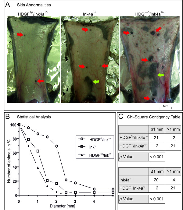
Results: Transgenic animals overexpressed HDGF in hair follicle melanocytes. Interestingly, primary melanocytes isolated from transgenic animals were not able to differentiate in vitro whereas cells isolated from wild type and HDGF-deficient animals were. Although, HDGF-/-/Ink4a+/- mice displayed an increased number of epidermoid cysts after exposure to UV light, no melanomas or premelanocytic alterations could be detected in this mouse model.

Conclusions: The results therefore provide no evidence that HDGF has a transforming capacity in tumor development. Our results in combination with previous findings point to a possible role in cell differentiation and suggest that HDGF promotes tumor progression after secondary upregulation and may represent another protein fitting into the concept of non-oncogene addiction of tumor tissue.


Figures

Figure 1
Figure 1
Generation of HDGFTyr transgenic mice. A: Schematic map of the Tyrosinase-HDGFgene construct. The Tyrosinase Promotor/Enhancer element is located approximately 6.1 kb in front of exonI. The HDGF gene consists of the complete exon/intron structure, except intronI was shortened from 6, 377 bp to 786 bp. The construct was separated from the vector backbone by incubation with the restriction enzyme BssHII, purified, and injected into the pronucleus of fertilized mouse egg cells. Boxes 1-6 indicate exons (white parts represent non-coding, black parts indicate coding regions). Arrows show the site of primer annealing for PCR-genotyping. The probe used for Southern blot analysis spans exonIV, intronIV and exonV. B: Examples of a polymerase chain reaction (PCR) genotyping of mouse tail DNA. Primers a and b were used to amplify the 943 bp fragment. Wildtype DNA was used as a negative control. 10 ng of Tyr-HDGFgene construct plasmid DNA was used as a positive control. C: Exemplary illustration of a Southern blot analysis of mouse tail DNA from the PCR-positive offspring 7159 and 889 and the PCR-negative offspring 7165 and wildtype mouse tail DNA as a control. The radioactively labeled probe spanning exonIV, intronIV and exonV hybridizes to both fragments of the wildtype HDGF-allele (4, 386 bp) and the Tyr-HDGFgene construct (2, 570 bp).
Figure 2
Figure 2
Detection of melanocyte-specific HDGF-overexpression. A: Immunofluorescence of skin samples from wildtype, HDGFTyr, HDGFTyr/HDGF-/- and HDGF-/- mice. Paraffin sections were stained with affinity-purified anti-mouse HDGF antibody. Bound antibody was detected by incubation with Cy3 conjugated secondary anti-rabbit antibody. Nuclei were stained with DAPI. a-e HDGF; f-j merge of HDGF, DAPI and transmitted light. Green arrows point at HDGF positive melanocyte nuclei. Scale bar equates 100 μm. B: Primary melanocyte cell culture. Melanocytes were isolated from newborn HDGFTyr, wildtype and HDGF-/- mice. After 2-3 weeks melanocytes from wildtype and HDGF-/- mice grew in a sub-confluent cell layer, whereas very few melanocytes from HDGFTyr animals (red arrows) were able to survive in cell culture. a-c 100 × magnification; d-f 400 × magnification. Scale bar equates 100 μm. C: Western blot analysis of melanocyte lysates. a HDGF-level of primary melanocytes from HDGFTyr, wildtype and HDGF-/- mice was compared to the level of complete skin and B16F10 (murine melanoma cell line) lysates. 10 μg total protein was loaded onto an SDS-gel. b Equal loading of melanocyte lysate was assessed by staining the membrane with Coomassie solution.
Figure 3
Figure 3
Statistical analysis of pigmented skin abnormalities (PSA). A: Overview of the inner side of skins from HDGFTyr/Ink4a+/-, Ink4a+/-, and HDGF-/-/Ink4a+/- mice showing PSA. Scale bar equates 1 cm. B: Statistical analysis of the occurrence of (PSA) of the different genotypes. HDGFTyr/Ink4a+/- n = 23, Ink4a+/- n = 24; HDGF-/-/Ink4a+/- n = 23. C: Chi-square contingency table was applied for the category: largest PSA was ≤ 1mm or > 1mm. Contingency tables were carried out for the following pairs of genotypes: HDGFTyr/Ink4a+/- - HDGF-/-/Ink4a+/- and Ink4a+/- - HDGF-/-/Ink4a+/-. Analysis was regarded significant for p < 0.01.
Figure 4
Figure 4
Histological analysis of pigmented skin abnormalities (PSA). A: Serial sections of PSA were either directly HE stained (a-c) or bleached in a benzylalcohol/aceton/H2O2 based bleaching solution before HE-staining was performed (d-f). Yellow arrows point to the pigmented and to the bleached areas respectively within the cyst lumen; green arrows point to pigmented and bleached melanocytes respectively. B: Immunofluorescence of epidermoid cysts. HDGF was detected by incubation with anti-mouse HDGF antibody and Cy3-conjugated goat-anti-rabbit antibody (a-f). Nuclei were stained with DAPI. a-c HDGF; d-f merge of HDGF, DAPI and transmitted light. Green arrows point to HDGF-positive nuclei in pigmented areas. Scale bars equate 100 μm.

Similar articles

Cited by

References

    1. Nakamura H, Izumoto Y, Kambe H, Kuroda T, Mori T, Kawamura K, Yamamoto H, Kishimoto T. Molecular cloning of complementary DNA for a novel human hepatoma-derived growth factor. Its homology with high mobility group-1 protein. J Biol Chem. 1994;269(40):25143–25149. - PubMed
    1. Nakamura H, Kambe H, Egawa T, Kimura Y, Ito H, Hayashi E, Yamamoto H, Sato J, Kishimoto S. Partial purification and characterization of human hepatoma-derived growth factor. Clin Chim Acta. 1989;183(3):273–284. doi: 10.1016/0009-8981(89)90361-6. - DOI - PubMed
    1. Abouzied MM, El-tahir HM, Prenner L, Haberlein H, Gieselmann V, Franken S. Hepatoma-derived Growth Factor:Significance of Amino Acid Residues 81-100 in Cell Surface Interaction and Proliferative Activity. J Biol Chem. 2005;280(12):10945–10954. doi: 10.1074/jbc.M414652200. - DOI - PubMed
    1. Everett AD, Bushweller J. Hepatoma derived growth factor is a nuclear targeted mitogen. Curr Drug Targets. 2003;4(5):367–371. doi: 10.2174/1389450033490975. - DOI - PubMed
    1. Everett AD, Stoops T, McNamara CA. Nuclear targeting is required for hepatoma-derived growth factor-stimulated mitogenesis in vascular smooth muscle cells. J Biol Chem. 2001;276(40):37564–37568. doi: 10.1074/jbc.M105109200. - DOI - PubMed

Substances

LinkOut - more resources