

Cattle remain immunocompetent during the acute phase of foot-and-mouth disease virus infection
- PMID: 22014145
- PMCID: PMC3207891
- DOI: 10.1186/1297-9716-42-108
Cattle remain immunocompetent during the acute phase of foot-and-mouth disease virus infection
Abstract
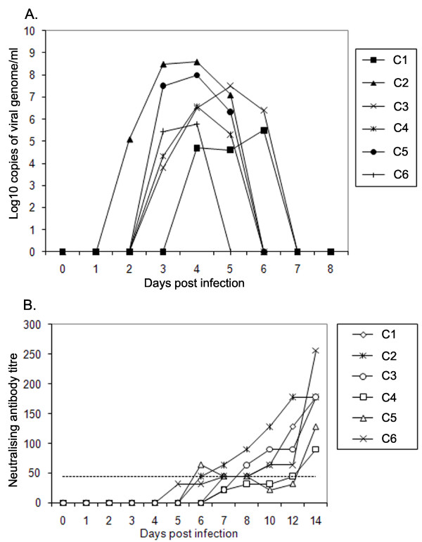
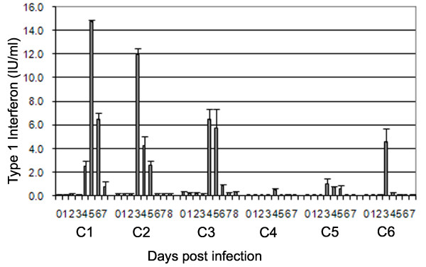
Infection of cattle with foot-and-mouth disease virus (FMDV) results in the development of long-term protective antibody responses. In contrast, inactivated antigen vaccines fail to induce long-term protective immunity. Differences between susceptible species have also been observed during infection with FMDV, with cattle often developing persistent infections whilst pigs develop more severe symptoms and excrete higher levels of virus. This study examined the early immune response to FMDV in naïve cattle after in-contact challenge. Cattle exposed to FMDV were found to be viraemic and produced neutralising antibody, consistent with previous reports. In contrast to previous studies in pigs these cattle did not develop leucopenia, and the proliferative responses of peripheral blood mononuclear cells to either mitogen or third party antigen were not suppressed. Low levels of type 1 interferon and IL-10 were detected in the circulation. Taken together, these results suggest that there was no generalised immunosuppression during the acute phase of FMDV infection in cattle.
Figures

Similar articles
-
Cell mediated innate responses of cattle and swine are diverse during foot-and-mouth disease virus (FMDV) infection: a unique landscape of innate immunity.Immunol Lett. 2013 May;152(2):135-43. doi: 10.1016/j.imlet.2013.05.007. Epub 2013 May 30. Immunol Lett. 2013. PMID: 23727070 Free PMC article. Review.
-
Foot-and-mouth disease virus exhibits an altered tropism in the presence of specific immunoglobulins, enabling productive infection and killing of dendritic cells.J Virol. 2011 Mar;85(5):2212-23. doi: 10.1128/JVI.02180-10. Epub 2010 Dec 22. J Virol. 2011. PMID: 21177807 Free PMC article.
-
Systemic immune response and virus persistence after foot-and-mouth disease virus infection of naïve cattle and cattle vaccinated with a homologous adenovirus-vectored vaccine.BMC Vet Res. 2016 Sep 15;12:205. doi: 10.1186/s12917-016-0838-x. BMC Vet Res. 2016. PMID: 27634113 Free PMC article.
-
Foot-and-mouth disease virus virulence in cattle is co-determined by viral replication dynamics and route of infection.Virology. 2014 Mar;452-453:12-22. doi: 10.1016/j.virol.2014.01.001. Epub 2014 Jan 25. Virology. 2014. PMID: 24606678
-
Virus-Host Interactions in Foot-and-Mouth Disease Virus Infection.Front Immunol. 2021 Feb 26;12:571509. doi: 10.3389/fimmu.2021.571509. eCollection 2021. Front Immunol. 2021. PMID: 33717061 Free PMC article. Review.
Cited by
-
Virulence and Immune Evasion Strategies of FMDV: Implications for Vaccine Design.Vaccines (Basel). 2024 Sep 19;12(9):1071. doi: 10.3390/vaccines12091071. Vaccines (Basel). 2024. PMID: 39340101 Free PMC article. Review.
-
The pathogenesis of foot-and-mouth disease virus: current understandings and knowledge gaps.Vet Res. 2025 Jun 16;56(1):119. doi: 10.1186/s13567-025-01545-5. Vet Res. 2025. PMID: 40524230 Free PMC article. Review.
-
Cell mediated innate responses of cattle and swine are diverse during foot-and-mouth disease virus (FMDV) infection: a unique landscape of innate immunity.Immunol Lett. 2013 May;152(2):135-43. doi: 10.1016/j.imlet.2013.05.007. Epub 2013 May 30. Immunol Lett. 2013. PMID: 23727070 Free PMC article. Review.
-
A Comprehensive Review of the Immunological Response against Foot-and-Mouth Disease Virus Infection and Its Evasion Mechanisms.Vaccines (Basel). 2020 Dec 14;8(4):764. doi: 10.3390/vaccines8040764. Vaccines (Basel). 2020. PMID: 33327628 Free PMC article. Review.
-
Laboratory animal models to study foot-and-mouth disease: a review with emphasis on natural and vaccine-induced immunity.J Gen Virol. 2014 Nov;95(Pt 11):2329-2345. doi: 10.1099/vir.0.068270-0. Epub 2014 Jul 7. J Gen Virol. 2014. PMID: 25000962 Free PMC article. Review.
References
-
- Paton DJ, King DP, Knowles NJ, Hammond J. Recent spread of foot-and-mouth disease in the Far East. Vet Rec. 2010;166:569–570. - PubMed
Publication types
MeSH terms
Substances
Grants and funding
LinkOut - more resources
Full Text Sources
