Comparative Study
. 2011 Oct 20;12(1):139.
doi: 10.1186/1465-9921-12-139.

Activated MCTC mast cells infiltrate diseased lung areas in cystic fibrosis and idiopathic pulmonary fibrosis

Affiliations
Comparative Study

Activated MCTC mast cells infiltrate diseased lung areas in cystic fibrosis and idiopathic pulmonary fibrosis

Cecilia K Andersson et al. Respir Res. .

Abstract

Background: Although mast cells are regarded as important regulators of inflammation and tissue remodelling, their role in cystic fibrosis (CF) and idiopathic pulmonary fibrosis (IPF) has remained less studied. This study investigates the densities and phenotypes of mast cell populations in multiple lung compartments from patients with CF, IPF and never smoking controls.

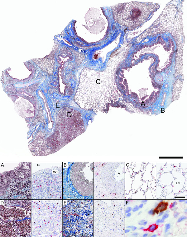
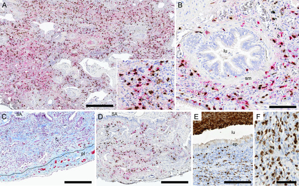
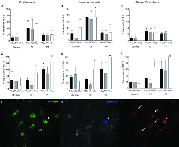
Methods: Small airways, pulmonary vessels, and lung parenchyma were subjected to detailed immunohistochemical analyses using lungs from patients with CF (20 lung regions; 5 patients), IPF (21 regions; 7 patients) and controls (16 regions; 8 subjects). In each compartment the densities and distribution of MCT and MCTC mast cell populations were studied as well as the mast cell expression of IL-6 and TGF-β.

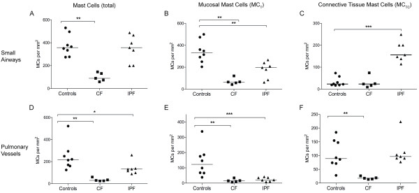
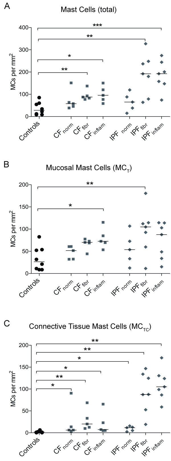
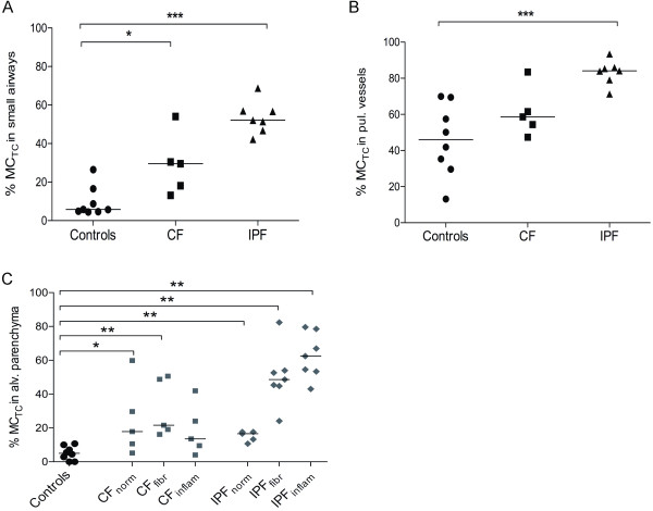
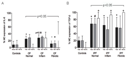
Results: In the alveolar parenchyma in lungs from patients with CF, MCTC numbers increased in areas showing cellular inflammation or fibrosis compared to controls. Apart from an altered balance between MCTC and MCT cells, mast cell in CF lungs showed elevated expression of IL-6. In CF, a decrease in total mast cell numbers was observed in small airways and pulmonary vessels. In patients with IPF, a significantly elevated MCTC density was present in fibrotic areas of the alveolar parenchyma with increased mast cell expression of TGF-β. The total mast cell density was unchanged in small airways and decreased in pulmonary vessels in IPF. Both the density, as well as the percentage, of MCTC correlated positively with the degree of fibrosis. The increased density of MCTC, as well as MCTC expression of TGF-β, correlated negatively with patient lung function.

Conclusions: The present study reveals that altered mast cell populations, with increased numbers of MCTC in diseased alveolar parenchyma, represents a significant component of the histopathology in CF and IPF. The mast cell alterations correlated to the degree of tissue remodelling and to lung function parameters. Further investigations of mast cells in these diseases may open for new therapeutic strategies.


Figures

Figure 1
Figure 1
Representative low magnification overview of a lung section from a patient with CF, stained with Masson's trichrome staining. Panel A-E show representative micrographs in different anatomical compartments of the lung from a patient with CF: (A) small airway, (B) pulmonary vessel, (C) normal alveolar parenchyma, (D) region of inflammation in alveolar parenchyma and (E) region of fibrosis in alveolar parenchyma. Consecutive sections were used for immunohistochemical double staining of tryptase-positive mast cells (MCT: permanent red) and chymase-positive mast cells (MCTC: DAB- brown) shown to the right in each panel. F represents a close-up image (600×) of neighbouring MCTC and MCT cells. Scale bars: Overview = 2000 μm, A-E = 100 μm. lu: airway lumen; ep: small airway epithelium; v: pulmonary vessel; and alv: alveolar parenchyma.
Figure 2
Figure 2
Representative micrographs from a patient with IPF. Panels A-B show lung section stained with immunohistochemical double staining for tryptase-positive mast cells (MCT: permanent red) and chymase-positive mast cells (MCTC: DAB- brown): alveolar parenchyma with interstitial fibrosis (A) and small airway (B). Picture C shows alveolar parenchyma with interstitial fibrosis and pulmonary pleura. High density of collagen is seen in blue with trichrome staining and picture D shows the corresponding area from a consecutive section double stained for MCT and MCTC. In picture E lung tissue from a patient with CF is shown. High density of MPO+ neutrophils is seen in the subepithelial tissue, in the airway lumen (E), and in the alveolar parenchyma (F). Scale bars: A, C-D = 300 μm, B and E = 100 μm and F = 50 μm. lu: airway lumen; ep: small airway epithelium; v: pulmonary vessel; pl: pleura and alv: alveolar parenchyma.
Figure 3
Figure 3
Total mast cell densities (A, D) and densities of each subtype, MCT (B, E) and MCTC (C, F), in lung tissue compartments of never-smoking controls and patients with CF and IPF. Data are presented as mast cells per mm2 lung tissue and presented for small airways (A-C) and pulmonary vessels (D-F). Horizontal line denotes median value. Data are expressed as scatter plots where horizontal lines denote median values. Statistical differences to never-smoking controls using Mann-Whitney where * denotes p < 0.05, and ** p < 0.01.
Figure 4
Figure 4
Mast cell densities in alveolar parenchyma from never-smoking controls and patients with CF and IPF. Data are presented as mast cells per mm2 lung tissue. The total mast cell densities (A) and densities of each subtype, MCT (B) and MCTC (C), are presented in regions of normal, inflammatory and fibrotic alveolar parenchyma in each disease. Data are expressed as scatter plots where horizontal lines denote median values. Statistical differences to never-smoking controls using Mann-Whitney where * denotes p < 0.05, ** < 0.01, and *** p < 0.001.
Figure 5
Figure 5
The proportion of MCTC and MCT, expressed as the percentage of MCTC, in anatomical lung compartments in never-smoking controls and patients with CF and IPF. Data are presented for small airways (A), pulmonary vessels (B), and alveolar parenchyma (C). Data are presented as scatter plots, where horizontal lines denote median values. Statistical differences to never-smoking controls were analysed using Mann-Whitney and asterisks show significant difference where * denotes p < 0.05, ** < 0.01, and *** p < 0.001.
Figure 6
Figure 6
Expression patterns of interleukin (IL)-6 (A-C), and TGF-β (D-F) in small airways, pulmonary vessels, and alveolar parenchyma. In each graph, results are shown as the percentage of total mast cells, and of MCTC and MCT subtypes that are positive for the respective mediator. Data are expressed as medians with interquartile ranges. Statistical differences to never-smoking controls were analysed using Mann-Whitney and asterisks show significant difference where * denotes p < 0.05, ** < 0.01, and *** p < 0.001. Representative micrographs of triple staining of mast cell subtypes and TGF-β (G-I). Arrows indicate tryptase and TGF-β co-localisation and * indicates co-localisation of tryptase, chymase and TGF-β.
Figure 7
Figure 7
Mast cell expression of IL-6 in parenchyma in patients with CF (A), and mast cell expression of TGF-β in parenchyma in IPF patients (B). Overall significance for total mast cell expression in different parenchyma is shown in each picture. Significance to healthy controls is shown as: p < 0.05 for total mast cell expression (*), as well as MCT (#) and MCTC (+), and to normal CF parenchyma as: p < 0.05 for total mast cell expression (×) and MCT (●).

Similar articles

Cited by

References

    1. Nilsson G, Costa J, Metcalfe D. Mast cells and basophils. 3. Philadelphia: Lippincott Williams and Wilkins; 1999.
    1. Garbuzenko E, Berkman N, Puxeddu I, Kramer M, Nagler A, Levi-Schaffer F. Mast cells induce activation of human lung fibroblasts in vitro. Exp Lung Res. 2004;30:705–721. doi: 10.1080/01902140490517809. - DOI - PubMed
    1. Levi-Schaffer F, Rubinchik E. Mast cell/fibroblast interactions. Clin Exp Allergy. 1994;24:1016–1021. doi: 10.1111/j.1365-2222.1994.tb02737.x. - DOI - PubMed
    1. Rubinchik E, Levi-Schaffer F. Mast cells and fibroblasts: two interacting cells. Int J Clin Lab Res. 1994;24:139–142. doi: 10.1007/BF02592443. - DOI - PubMed
    1. Batlle M, Perez-Villa F, Lazaro A, Garcia-Pras E, Ramirez J, Ortiz J, Orus J, Roque M, Heras M, Roig E. Correlation between mast cell density and myocardial fibrosis in congestive heart failure patients. Transplant Proc. 2007;39:2347–2349. doi: 10.1016/j.transproceed.2007.06.047. - DOI - PubMed

Publication types

MeSH terms